Download

1 / 40
720 likes | 2.85k Views
Widal test. Widal test. Introduction It is a serological approach for the diagnosis of typhoid and paratyphoid fever in clinical laboratory. This is a test for the measurement of H and O agglutinins of typhoid and paratyphoid bacilli in patient’s sera. Widal test. Materials

E N D
Widal test Introduction • It is a serological approach for the diagnosis of typhoid and paratyphoid fever in clinical laboratory. • This is a test for the measurement of H and O agglutinins of typhoid and paratyphoid bacilli in patient’s sera.
Widal test Materials 1. Sera from suspected patients 2. Antigens Suspension of S. typhi "O" antigen, TO Suspension of S. typhi "H" antigen; TH Suspension of S. paratyphi A "H" antigen, PAH Suspension of S. schottmuelleri "H" antigen, PBH Suspension of S. hirschfeldii "H" antigen, PCH 3. Normal saline(0.9% NaCl) 4. Test tubes and pipettes
Widal test Procedures
↘ ↘ ↘ ↘ ↘ Widal test—two folds serial dilution of the serum Tubes 1 2 3 4 5 6 NS (ml) 0.5 0.5 0.5 0.5 0.5 0.5 Serum (ml) 0.5 0.5 0.5 0.5 0.5 0.5ml discarded (1:10)1:20 1:40 1:80 1:160 1:320 Antigen (ml) 0.5 0.5 0.5 0.5 0.5 0.5 dilution1:40 1:80 1:160 1:320 1:640 overnight in a 37℃ waterbath negative control
observation of the results Widal test • H agglutination: loose clump, like cotton or wool • O agglutination: compact film, scattering at the bottom of the tubes
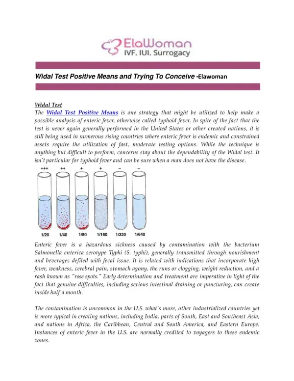
Widal test • Agglutination titer is determined as the highest dilution of serum which can cause ++bacteria agglutination. 1 2 3 4 5 6 TO ++++ ++ ++ + - - TH ++++ +++ ++ ++ - - PAH ++ + - - - - PBH - - - - - - PCH - - - - - - 1:40 1:80 1:160 1:320 1:640 Negative control
Widal test Interpretation of results
Experiment Isolation and Identification of Enterobacteriaceae
The common procedures of the Isolation and Identification of Enterobacteriaceae Specimen (feces or rectal swab) ↓ Differential or selective culture media (such as EMB agar) ↓ Colonies Lactose (-) lactose (+) ↓ Double sugar iron slant Other biochemical reactions slide agglutination tests (Such as urea slant)
5 students/group • Materials and Methods • Bacterial strains Slant culture of E.coli Slant culture of Proteusvulgaris Slant culture of S.typhi Slant culture of S.dysenteriae Slant culture of Enterobacter.aerogen
Culture medium and inoculation methods Note: Each bacterial strain will be inoculated into all the five kinds of culture media. After inoculation, incubate the plates and tubes overnight at 37℃.
Four-area streak dilution technique 1/10 1/5 1/4 EMB agar plate
Culture medium and inoculation methods Note: Each bacterial strain will be inoculated into all the five kinds of culture media. After inoculation, incubate the plates and tubes overnight at 37℃.
Culture medium and inoculation methods Note: Each bacterial strain will be inoculated into all the five kinds of culture media. After inoculation, incubate the plates and tubes overnight at 37℃.
Culture medium and inoculation methods Note: Each bacterial strain will be inoculated into all the five kinds of culture media. After inoculation, incubate the plates and tubes overnight at 37℃.
5 students/group (lab report) 1 2 3 4 5 E.coli Proteus S.dysenteriae E.aerogen S.typhi Double sugar iron Indole broth Urea slant Citrate slant EMB
Observe the results (1) Eosin-Methylene Blue Agar (EMB) Reagents: Eosin and Methylene-blue Carbohydrate Source: Lactose Principle: a precipitate can be formed at acidic pH Purpose: differentiate lactose fermenters from non-lactose fermenters. Interpretation: Lactose fermenter: purple Lactose non-fermenter: colorless
EMB agar E.coli: black and typically have a metallic green sheen negative
Observe the results • (2) Idole Broth purpose:Distinguish Enterobacteriaseae based on the ability to produce indole from tryptophan. principle: Interpretation • Positive Test - red ring • Negative Test - no color development Kovac’s reagent tryptophanase indole tryptophan rosindole (red)
Idole Broth negative positive
(3) Double suger iron slant main component: Glucose: 0.1% Lactose: 1% Phenol red: as an indicator Ferrous sulfate(FeSO4) :FeSO4+H2S →FeS(black)+H2SO4 purpose:It provides information about carbohydrate fermentation. Principle:
Only glucose fermenting Slant: oxidized →neutral →red Butt: not oxidized →yellow
Lactose fermenting yellow both on the slant and in the butt
H2S production FeSO4+H2S FeS(black)+H2SO4
Observe the results • (4) Citrate Slant purpose: Determine if organism can use citrate as its sole metabolic (carbon) energy source. reagents:bromothymol blue (a pH indicator) sodium citrate (the sole source of carbon) principle: sodium citrate sodium carbonate • Interpretation: • Positive: blue • Negative: green citrase
Observe the results • (5) Urea slant purpose: determine if organism can break down urea or not. reagents:urea, phenol red principle:if urease is present, urea is broken down to ammonia causing rise to pH • Interpretation: • Positive: red • Negative: pale yellow
slide agglutination test Procedures 1) Divide the slide into two equal parts 2) Add one drop of NSon the left side, and then on the right side 3) Emulsify S.typhi in the left side,and S.dysenteriae in the right side. 4) Add one drop of diagnostic sera for S.typhi on the left side, and then on the right side 5) Gently shake the slide and observe the results