Download
1 / 35
360 likes | 394 Views
Gain insights into osteomyelitis, its etiology, pathogenesis, and clinical features. Learn about pyogenic osteomyelitis, tuberculosis osteomyelitis, and infectious arthritis.

E N D
MUSCULOSKELETAL BLOCKPathologyOSTEOMYELITIS and SEPTIC ARTHRITIS
Objectives • Understand the etiology, pathogenesis and clinical features of osteomyelitis. • Be familiar with some of the terminology used in bone infections like: sequestrum, involucrum, Brodie abscess and Pott’s disease. • Understand the clinicopathological features of tuberculous osteomyelitis and infective arthritis.
Osteomyelitis refers to inflammation of the bone and marrow and is usually the result of infection OSTEOMYELITISDefinition
OSTEOMYELITIS Etiology • All types of organisms, including viruses, parasites, fungi and bacteria can produce osteomyelitis. • The most common are infections caused by certain pyogenic bacteria and mycobacteria • Staphylococcus aureus is responsible for 80% to 90% the cases of pyogenic osteomyelitis
PYOGENIC OSTEOMYELITIS :Bacteria which are common in certain conditions • Neonates: Escherichia coli and group B streptococci. • Persons with sickle cell disease: Salmonella
PYOGENIC OSTEOMYELITIS :Bacteria which are common in certain conditions • Patients with genitourinary tract infections or with intravenous drug abusers: E.coli, Klebsiella and Pseudomonas • Direct spread during surgery or open fractures (secondary to bone trauma): Mixed bacterial infections, including anaerobes
PYOGENIC OSTEOMYELITIS Routes of infection • Hematogenous spread, most common. • Extension from a contiguous site. • Direct implantation.
PYOGENIC OSTEOMYELITISPathogenesis Stages : • Acute • Sub acute • Chronic.
Once in bone, the bacteria proliferate and induce an acute inflammatory reaction. • The entrapped bone undergoes necrosis within the first 48 hours, and the bacteria and inflammation spread within the shaft of the bone and may reach the periosteum.
In children the periosteum is loosely attached to the cortex; sizable subperiosteal abscesses may form • Lifting of the periosteum further impairs the blood supply to the affected region, and both the suppurative and the ischemic injury may cause segmental bone necrosis; the dead piece of bone is known as a sequestrum.
. Rupture of the periosteum leads to a soft-tissue abscess and the eventual formation of a draining sinus.
After the first week chronic inflammatory cells become more numerous and their release of cytokines stimulates osteoclastic bone resorption, ingrowth of fibrous tissue, and the deposition of reactive bone in the periphery. • the newly deposited bone is known as an involucrum . • Brodie abscess is a small intraosseous abscess that frequently involves the cortex
PYOGENIC OSTEOMYELITIS • Clinical Course: • Fever ,chills, malaise, marked to intense throbbing pain over the affected region and swelling Diagnosis; • Sign/symptoms. • X-ray:a lytic focus of bone surrounded by a zone of sclerosis • Blood cultures • biopsy
Treatment requires aggressive antibiotic therapy. Inadequate treatment of acute osteomyelitis may lead to chronic osteomyelitis which is notoriously difficult to manage. Surgical removal of bony tissue may be required.
Chronicity may develop with: • delay in diagnosis • extensive bone necrosis • abbreviated antibiotic therapy • inadequate surgical debridement, • weakened host defenses.
PYOGENIC OSTEOMYELITIS • Complications: • Pathologic fracture. • Secondary amyloidosis • Endocarditis • Sepsis • Squamous cell carcinoma if the infection creates a sinus tract. • Rarely sarcoma in the affected bone
Tuberculousosteomyelitis Routes of entry; • Usually blood borne and originate from a focus of active visceral disease. • Direct extension (e.g. from a pulmonary focus into a rib or from tracheobronchial nodes into adjacent vertebrae) or spread via draining lymphatics.
Tuberculousosteomyelitis • The most common sites of skeletal involvement are: • thoracic and lumber vertebrae followed by the knees and hips • Pott’s disease is the involvement of spine • In patients with AIDS frequently multifocal
The infection breaks through the intervertebral discs and extends into the soft tissues forming abscesses. Pott’s disease
In Pott’s disease, the infection may breaks through the intervertebral discs and extends into the muscle forming Psoas abscesses
Tuberculousosteomyelitis • Histopathology: collections of epithelioid histiocytes and lymphocytes with caseation necrosis ZiehlNeelsen stain
Tuberculousosteomyelitis Clinical features : • Pain • Fever • Weight loss • May form an inguinal mass “ psoas abscess”.
Tuberculousosteomyelitis Complications • Bone destruction • Tuberculous arthritis • Sinus tract formation • Amyloidosis
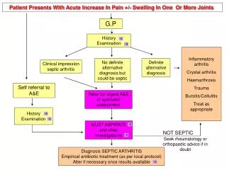
Infectious Arthritis • Microorganisms of all types can seed joints during hematogenous dissemination. • Articular structures can also become infected by direct inoculation or from contiguous spread from a soft-tissue abscess or focus of osteomyelitis. • Infectious arthritis is potentially serious, because it can cause rapid destruction of the joint and produce permanent deformities
Routes of infection: • Hematogenous • Contiguous spread from osteomyelitis • Contiguous spread from a soft tissue abscess • Iatrogenic • Traumatic
Infectious Arthritis-bacterial • Bacterial infections almost always cause an acute suppurative arthritis • Any bacteria can be causal: • Haemophilusinfluenzae predominates in children under age 2 years • S. aureus is the main causative agent in older children and adults • gonococcus is prevalent during late adolescence and young adulthood. • Individuals with sickle cell disease are prone to infection with Salmonella at any age.
Risk factors • Immune deficiencies (congenital and acquired) • Debilitating illness • Joint trauma • Chronic arthritis of any cause • Intravenous drug abuse
Infectious Arthritis • The infection involves only a single joint • usually the knee-followed in order by hip, shoulder, elbow, wrist, and sternoclavicular joints. • Joint aspiration is typically purulent • Culture allows identification of the causal agent.
Infectious Arthritis Clinical features: • sudden onset of pain • redness, and swelling of the joint with restricted range of motion. • Fever, leukocytosis, and elevated erythrocyte sedimentation rate
Infectious arthritis must be rapidly diagnosed and treated promptly to prevent irreversible and permanent joint damage.
Complication • Septic arthritis can lead to ankylosis and even fatal septicemia. • However, prompt antibiotic therapy and joint aspiration or drainage cures most patients.