Download
1 / 47
500 likes | 1.57k Views
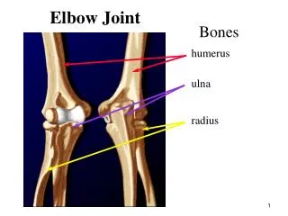
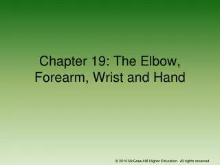
Chapter 23: The Elbow. Anatomy of the Elbow. Figure 23-1. Figure 23-2. Figure 23-3. Figure 23-4 A-C. Functional Anatomy. Complex that allows for flexion, extension, pronation and supination 145 degrees of flexion and 90 degrees of supination and pronation

E N D
Chapter 23: The Elbow © 2011 McGraw-Hill Higher Education. All rights reserved.
Anatomy of the Elbow © 2011 McGraw-Hill Higher Education. All rights reserved.
Figure 23-1 © 2011 McGraw-Hill Higher Education. All rights reserved.
Figure 23-2 © 2011 McGraw-Hill Higher Education. All rights reserved.
Figure 23-3 © 2011 McGraw-Hill Higher Education. All rights reserved.
Figure 23-4 A-C © 2011 McGraw-Hill Higher Education. All rights reserved.
Functional Anatomy Complex that allows for flexion, extension, pronation and supination 145 degrees of flexion and 90 degrees of supination and pronation Bony limitations, ligamentous support and muscular stability at the elbow help to protect it from overuse and traumatic injuries Elbow demonstrates a carrying angle due to distal projection of humerus Normal in females is 10-15 degrees, males 5 degrees Critical link in kinetic chain of upper extremity © 2011 McGraw-Hill Higher Education. All rights reserved.
Assessment of the Elbow History Past history Mechanism of injury When and where does it hurt? Motions that increase or decrease pain Type of, quality of, duration of, pain? Sounds or feelings? How long were you disabled? Swelling? Previous treatments? © 2011 McGraw-Hill Higher Education. All rights reserved.
Observations Deformities and swelling? Carrying angle Cubitus valgus versus cubitus varus Flexion and extension Cubitus recurvatum Elbow at 45 degrees Isosceles triangle (olecranon and epicondyles) Figure 23-5 Figure 23-8 Figure 23-7 © 2011 McGraw-Hill Higher Education. All rights reserved.
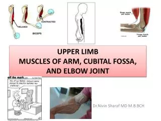
Palpation: Bony and Soft Tissue Humerus Medial and lateral epicondyles Olecranon process Radial head Radius Ulna Medial and lateral collateral ligaments Annular ligament Biceps brachii Brachialis Brachioradialis Pronator teres Triceps Supinator Wrist flexors and extensors © 2011 McGraw-Hill Higher Education. All rights reserved.
Special Tests Circulatory and Neurological Function Pulse should be taken at brachial artery and radial artery Skin sensation should be checked - determine presence of nerve root compression or irritation in cervical or shoulder region Tinel’s sign Ulnar nerve test Tap on ulnar nerve (in ulnar groove) Positive test is found when athlete complains of sensation along the forearm and hand Test for Capsular Injury Tested after hyperextension of elbow Elbow is flexed to 45 degrees, wrist is fully flexed and extended If joint pain is severe, moderate/severe sprain or fracture should be suspected (chronic injury may present same) © 2011 McGraw-Hill Higher Education. All rights reserved.
Valgus/Varus Stress Test Assess injury to the medial and lateral collateral ligaments, respectively Looking for gapping or complaint of pain Figure 23-10 © 2011 McGraw-Hill Higher Education. All rights reserved.
Medial and Lateral Epicondylitis Tests Elbow flexed to 45 degrees and wrist extension or flexion is resisted Pain at lateral or medial epicondyle, respectively indicates a positive test Pinch Grip Test Pinch thumb and index finger together Inability to touch fingers together indicates entrapment of anterior interosseous nerve between heads of pronator muscle Pronator Teres Syndrome Test Forearm pronation is resisted Increased pain proximally over pronator teres indicates a positive test © 2011 McGraw-Hill Higher Education. All rights reserved.
Figure 23-11 & 12 © 2011 McGraw-Hill Higher Education. All rights reserved.
Functional Evaluation Pain and weakness are evaluated through AROM, PROM and RROM Flexion, extension, pronation and supination ROM of pronation and supination are particularly noted Figure 23-13 & 14 © 2011 McGraw-Hill Higher Education. All rights reserved.
Recognition and Management of Injuries to the Elbow Subject to injury due to broad range of motion, weak lateral bone structure, and relative exposure to soft tissue damage Many activities place excessive stress on joint Locking motion of some activities, use of implements, and involvement in throwing motion make elbow extremely susceptible © 2011 McGraw-Hill Higher Education. All rights reserved.
Contusion Etiology Vulnerable area due to lack of padding Result of direct blow or repetitive blows Signs and Symptoms Swelling (rapidly after irritation of bursa or synovial membrane) Management Treat w/ RICE immediately for at least 24 hours If severe, refer for X-ray to determine presence of fracture © 2011 McGraw-Hill Higher Education. All rights reserved.
Olecranon Bursitis Etiology Superficial location makes it extremely susceptible to injury (acute or chronic) --direct blow Signs and Symptoms Pain, swelling, and point tenderness Swelling will appear almost spontaneously and w/out usual pain and heat Figure 23-17 © 2011 McGraw-Hill Higher Education. All rights reserved.
Management In acute conditions, compression for at least 1 hour Chronic cases require superficial therapy primarily involving compression If swelling fails to resolve, aspiration may be necessary Can be padded in order to return to competition © 2011 McGraw-Hill Higher Education. All rights reserved.
Muscle Strains Etiology MOI is excessive resistive motion (falling on outstretched arm), repeated microtears that cause chronic injury Rupture of distal biceps is most common muscle rupture of the upper extremity Signs and Symptoms Active or resistive motion produces pain; point tenderness in muscle, tendon, or lower part of muscle belly Management RICE and sling in severe cases Follow-up w/ cryotherapy, ultrasound and exercise If severe loss of function encountered - should be referred for X-ray (rule out avulsion or epiphyseal fx) © 2011 McGraw-Hill Higher Education. All rights reserved.
Ulnar Collateral Ligament Injuries Etiology Injured as the result of a valgus force from repetitive trauma Can also result in ulnar nerve inflammation, or wrist flexor tendinitis; overuse flexor/pronator strain, ligamentous sprains; elbow flexion contractures or increased instability Signs and Symptoms Pain along medial aspect of elbow; tenderness over MCL Associated paresthesia, positive Tinel’s sign Pain w/ valgus stress test at 20 degrees; possible end-point laxity X-ray may show hypertrophy of humeral condyle, posteromedial aspect of olecranon, marginal osteophytes; calcification w/in MCL; loose bodies in posterior compartment © 2011 McGraw-Hill Higher Education. All rights reserved.
Ulnar Collateral Ligament Injuries (cont.) Management Conservative treatment begins w/ RICE and NSAID’s W/ resolution, strengthening should be performed; analysis of the throwing motion (if applicable) Surgical intervention may be necessary (Tommy John procedure) Involves reconstruction using palmaris longus autograft and occasionally transposition of the ulnar nerve Throwing athlete can return to activity 22-26 weeks post surgery with full-recovery taking 18-24 months © 2011 McGraw-Hill Higher Education. All rights reserved.
Lateral Epicondylitis (Tennis Elbow) Etiology Repetitive microtrauma to insertion of extensor muscles of lateral epicondyle Tendinosis with degeneration of tendon without inflammation Signs and Symptoms Aching pain in region of lateral epicondyle after activity Pain worsens and weakness in wrist and hand develop Elbow has decreased ROM; pain w/ resistive wrist extension © 2011 McGraw-Hill Higher Education. All rights reserved.
Lateral Epicondylitis (continued) Management RICE, NSAID’s and analgesics ROM exercises and PRE, deep friction massage, hand grasping while in supination, avoidance of pronation motions Mobilization and stretching in pain free ranges Use of a counter force or neoprene sleeve Mechanics and skills training in order to avoid recurrence Figure 23-19 © 2011 McGraw-Hill Higher Education. All rights reserved.
Medial Epicondylitis Etiology Repeated forceful flexion of wrist and extreme valgus torque of elbow May involve pronator teres, flexor carpi radialis and ulnaris, and palmaris longus tendons Can be associated with ulnar nerve neuropathy Signs and Symptoms Pain produced w/ forceful flexion or extension Point tenderness and mild swelling Passive movement of wrist seldom elicits pain, but active movement does © 2011 McGraw-Hill Higher Education. All rights reserved.
Management Sling, rest, cryotherapy or heat through ultrasound Analgesic and NSAID's Curvilinear brace below elbow to reduce elbow stressing Severe cases may require splinting and complete rest for 7-10 days © 2011 McGraw-Hill Higher Education. All rights reserved.
Elbow Osteochondritis Dissecans Etiology Impairment of blood supply to anterior surface resulting in degeneration of articular cartilage, creating loose bodies Repetitive microtrauma in movements of elbow rotation, extension, valgus stress causing compression of the radial head and shearing of the radiocapitellar joint Seen in young athletes involved in throwing motion Panner’s disease in incidents of children age <10 Osteochondrosis of capitellum due to localized avascular necrosis Signs and Symptoms Sudden pain, locking; range usually returns in a few days © 2011 McGraw-Hill Higher Education. All rights reserved.
Signs and Symptoms (continued) Swelling, pain at radiohumeral joint, crepitus, decreased ROM (full extension); grating w/ pronation and supination X-ray may show flattening and crater of capitulum w/ loose bodies Management Activity restriction for 6-12 weeks; NSAID’s Splint and cast applied for cases of extensive deterioration If repeated locking occurs, loose bodies are removed surgically © 2011 McGraw-Hill Higher Education. All rights reserved.
Little League Elbow Etiology Caused by repetitive microtraumas that occur from throwing (not type of pitch) May result in numerous disorders of growth in the pitching elbow Linked to: Accelerated apophyseal growth region and delay in medial epicondyle growth plate Traction apophysitis with possible fragmentation of medial epicondylar apophysis Avulsion of medial epicondyle or radial head Osteochondrosis of humeral capitellum Non-union stress fracture of olecranon epiphysis © 2011 McGraw-Hill Higher Education. All rights reserved.
Little League Elbow (continued) Signs and Symptoms Onset is slow; slight flexion contracture, including tight anterior joint capsule and weakness in triceps Patient may complain of locking or catching sensation Decreased ROM of forearm pronation and supination Management RICE, NSAID’s and analgesics Throwing stops until pain resolved and full ROM is regained Gentle stretching and triceps strengthening Throwing under supervision w/ good technique to prevent recurrence © 2011 McGraw-Hill Higher Education. All rights reserved.
Cubital Tunnel Syndrome Etiology Pronounced cubital valgus may cause deep friction problem Ulnar nerve dislocation Traction injury from valgus force, irregularities w/ tunnel, subluxation of ulnar nerve due to lax impingement, or progressive compression of ligament on the nerve Signs and Symptoms Pain medially which may be referred proximally or distally Tenderness in cubital tunnel on palpation and hyperflexion Intermittent paresthesia in 4th and 5th fingers © 2011 McGraw-Hill Higher Education. All rights reserved.
Cubital Tunnel Syndrome (continued) Management Rest, immobilization for 2 weeks w/ NSAID’s Splinting or surgical decompression or transposition of subluxating nerve may be necessary Patient must avoid hyperflexion and valgus stresses © 2011 McGraw-Hill Higher Education. All rights reserved.
Dislocation of the Elbow Etiology Caused by fall on outstretched hand w/ elbow extended or severe twist while flexed Bones can be displaced backward, forward, or laterally Distinguishable from fracture because lateral and medial epicondyles are normally aligned w/ shaft of humerus Signs and Symptoms Swelling, severe pain, disability Complications w/ median and radial nerves and blood vessels Often a radial head fracture is involved © 2011 McGraw-Hill Higher Education. All rights reserved.
Elbow Dislocation © 2011 McGraw-Hill Higher Education. All rights reserved.
Management Cold and pressure immediately w/ sling Refer for reduction Neurological and vascular fxn must be assessed prior to and following reduction Physician should reduce - immediately Immobilization following reduction in flexion for 3 weeks Hand grip and shoulder exercises should be used while immobilized Following initial healing, heat and passive exercise can be used to regain full ROM Massage and joint movement that are too strenuous should be avoided before complete healing due to high probability of myositis ossificans ROM and strengthening should be performed and initiated by patient (forced stretching should be avoided) © 2011 McGraw-Hill Higher Education. All rights reserved.
Fractures of the Elbow Etiology Fall on flexed elbow or from a direct blow Fracture can occur in any one or more of the bones Fall on outstretched hand often fractures humerus above condyles or between condyles Condylar fracture may result in gunstock deformity Direct blow to olecranon or radial head may result in fracture Signs and Symptoms May not result in visual deformity Hemorrhaging, swelling, muscle spasm © 2011 McGraw-Hill Higher Education. All rights reserved.
Elbow Fractures (continued) Management Decrease ROM, neurovascular status must be monitored Surgery is used to stabilize adult unstable fracture, followed by early ROM exercises Stable fractures do not require surgery Removable splints are used for 6-8 weeks Figure 23-22 © 2011 McGraw-Hill Higher Education. All rights reserved.
Volkmann’s Contracture Etiology Associate w/ humeral supracondylar fractures, causing muscle spasm, swelling, or bone pressure on brachial artery, inhibiting circulation to forearm Can become permanent There may be loss of motor and sensory function Classic case involves median nerve Results from insufficient arterial perfusion and venous stasis followed by ischemic degeneration Irreversible muscle damage occurs after 4-6 hours Edema further impairs circulation propagating muscle necrosis Necrosis may lead to secondary fibrosis and calcification © 2011 McGraw-Hill Higher Education. All rights reserved.
Signs and Symptoms Pain in forearm - increased w/ passive extension of fingers Pain is followed by cessation of brachial and radial pulses, coldness in arm Decreased motion Management Remove elastic wraps or casts Close monitoring must occur Figure 23-23 © 2011 McGraw-Hill Higher Education. All rights reserved.
Pronator Teres Syndrome • Etiology • Entrapment of median nerve • Proximal to the elbow joint • In the pronator teres muscle as the nerve passes between the superficial and deep heads of the muscle • May become trapped due to edema or muscle hypertrophy • Signs & Symptoms • Sensory deficits – numbness, tingling, pins & needles (digits 1-4) • Motor deficits – loss of flexion and opposition, weakness with pronation • Symptoms reproduced with tightly gripping and resisted pronation • Management • Rest, NSAID’s, TENS for pain, modified activity • Decompression surgery if treatment is unsuccessful © 2011 McGraw-Hill Higher Education. All rights reserved.
Rehabilitation of the Elbow General Body Conditioning Must maintain pre-injury fitness levels - cardiovascular and strength (lower body) Flexibility Restoring ROM is critical in elbow rehab Variety of approaches can be used as long as they don’t force the joint Figure 23-24 © 2011 McGraw-Hill Higher Education. All rights reserved.
Figure 23-25 A & B • Joint Mobilizations • Loss of proper arthrokinematics following immobilization is expected • Joint mobilization and traction can be very useful to increase mobility and decrease pain through restoration of accessory motions © 2011 McGraw-Hill Higher Education. All rights reserved.
Strengthening Achieved through low-resistance, high-repetition exercises - must be pain free Shoulder and grip exercises should also be performed Continuous passive motion units followed by dynamic splinting is ideal following surgery Isometrics can be used while elbow is immobilized PNF and isokinetics are useful in early and intermediate active stages of rehab A graded PRE program w/ tubing, weights or manual resistance should be included © 2011 McGraw-Hill Higher Education. All rights reserved.
Strengthening Exercises Plyometric Exercises Figure 23-26 Figure 23-27 © 2011 McGraw-Hill Higher Education. All rights reserved.
Closed Kinetic Chain Exercises Figure 23-28 • Closed kinetic chain activities should also be incorporated • Assist in both static and dynamic stability to the elbow • Proprioceptive training should also incorporated © 2011 McGraw-Hill Higher Education. All rights reserved.
Functional Progressions Will enhance healing and performance PNF, swimming, pulley machines and rubber tubing Simulate sports activities Should include steps Warm-up Gradual build up to activity, becoming increasingly more difficult © 2011 McGraw-Hill Higher Education. All rights reserved.
Return to Activity Can re-engage in activity when criteria has successfully been completed ROM w/in normal limits, strength should be equal w/ no complaint of pain Return should progress with use of restrictions in an effort to objectively measure activity progression © 2011 McGraw-Hill Higher Education. All rights reserved.