Download
1 / 52
520 likes | 749 Views
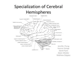
CH10. Cerebral hemispheres and vascular supply. By: Laurence Poliquin-Lasnier R2 Neurology. Outline. Review of the main functional cortical areas Anterior circulation Posterior circulation Circle of Willis Anatomy and vascular territories of: a) ACA b ) MCA c ) PCA

E N D
CH10. Cerebral hemispheres and vascular supply By: Laurence Poliquin-Lasnier R2 Neurology
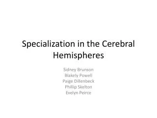
Outline • Review of the main functional cortical areas • Anterior circulation • Posterior circulation • Circle of Willis • Anatomy and vascular territories of: a) ACA b) MCA c) PCA • Clinical syndromes of the 3 main cerebral arteries • Venous drainage of the cerebral hemispheres • Clinical scenarios
4 segments of internal carotid artery • Cervical segment • Petrous segment • Cavernous segment • Intracranial/supraclinoid segment
The anterior circulation: branches of the supraclinoid/intracranial carotid artery • Mnemonic “OPAAM” • O = Ophtalmic artery • P = Posterior communicating artery • A = Anterior choroidal artery • A = Anterior cerebral artery • M = Middle cerebral artery
The anterior circulation: branches of the intracranial carotid artery
3 main arteries • Anterior cerebral artery (ACA) • Middle cerebral artery (MCA) • Posterior cerebral artery (PCA) • ACA and MCA arise from the internal carotid artery • PCA arise from the basilar artery
Vascular territories of the 3 main cerebral arteries • Vascular territories of the superficial cerebral structures • Vascular territories of the deep cerebral structures
Vascular territories of deep cerebral structures • Lenticulostriate arteries • Anterior choroidal artery • Recurrent artery of Heubner • Thalamoperforator arteries
Clinical syndromes of the 3 main cerebral arteries • MCA • ACA • PCA
Where is the lesion? • R face/arm UMN weakness, broca aphasia,+/- R face/arm cortical-type sensory loss
Where is the lesion? • R pure motor hemiparesis (UMN) • R hemiplegia, R hemianesthesia, R homonymous hemianopsia, global aphasia, L gaze preference
Where is the lesion? • R leg weakness (UMN), R leg cortical-type sensory loss, grasp, dishinibition • R homonymous hemianopia, alexia without agraphia
Clinical pearl: Alexia without agraphia • Lesion in dominant (usually L) occipital cortex extending to the posterior corpus callosum • Prevents processing of information in R visual field, including written material • Information about L visual field is transmitted to R occipital lobe, but cannot cross to the left to the language areas by the corpus callosum lesion
Lacunar syndromes • Lacune: small vessel infarct • Ressemble small lake or cavity when examined on pathologic section 6 major lacunar syndromes: • Pure motor hemiparesis • Pure sensory stroke • Ataxic hemiparesis • Sensorimotor stroke • Dysarthria-clumsy hand syndrome • Basal ganglia lacune
1- Pure motor hemiparesis • Unilateral face, arm, leg (UMN) weakness with dysarthria • Location: • Posterior limb internal capsule (common) • Lenticulostriate, anterior choroidal, thalamoperforator • Ventral pons (common) • Ventral penetrating branches of basilar artery • Corona radiata • Small MCA branches • Cerebral peduncle • Small MCA branches
2- Pure sensory stroke • Sensory loss to all primary modalities in the contralateral face and body • Location: • Ventral posterior lateral nucleus (VPL) of thalamus • Thalamoperforator branches of PCA
3- Ataxia hemiparesis • Pure motor hemiparesis with ataxia on same side as weakness • Location: Same as pure motor hemiparesis • Vascular supply: Same as pure motor hemiparesis
4- Sensorimotor (thalamocapsular) • Contralateral face/arm/leg sensory loss and weakness +/- dysarthria • Location: • Posterior limb internal capsule and either thalamic VPL or thalamic somatosensory radiations • Thalamoperforator arteries or lenticulostriate arteries
5- Dysarthria-clumsy hand • Facial weakness, dysarthria, dysphagia, and slight weakness and clumsiness of one hand • Location: • Pons • Pontine arteries • Genu of internal capsule
6- Basal ganglia lacune • Hemiballismus or asymptomatic • Locations: • Caudate, putamen, globuspallidus, or subthalamic nucleus • Lenticulostriate, anterior choroidal, thalamoperforator, or heubner’s arteries
Overview of venous drainage • Superficial veins drain into the superior sagittal sinus and cavernous sinus • Deep veins drain into great vein of Galen • Majority of veins ultimately drain to the internal jugular veins • Superior sagittal sinus –> transverse sinuses ->sigmoid sinus -> jugular foramen to become the internal jugular vein • Cavernous sinus (int carotid artery, CN III-IV-V-VI) ->superior petrosal sinus -> transverse sinus • Cavernous sinus -> inferior petrosal sinus ->internal jugular vein
Deep venous drainage • Internal cerebral veins, basal veins of Rosenthal, and other veins ->great cerebral vein of Galen -> joined by inferior sagittal sinus –> to form straight sinus • Confluence of sinus (torcularHerophili) = superior sagittal sinus + straight sinus + occipital sinus • Confluence of sinus drained by transverse sinus
Clinical scenario #1 • ID: 67yo woman • PMHx: HTN, PVD, smoker • HPI: after breakfast, she tried to stand up and suddenly felt she could not support her weight -> fell -> 911 • Physical: • Alert & oriented • Unaware at times of L sided weakness • Language fluent • CN normal except minimally decreased L nasolabial fold + mild dysarthria
Clinical scenario #1 • Motor: 5/5 except 1-2/5 in L leg prox and distal and 4/5 prox L arm • L leg hyperreflexia, L Babinski • Sensory: inconsistent decreased response to pinprick on L • Tactile extinction on L • One month later, partially recovered power, but feels that her L arm is out of control, grasp onto things without her being aware and would have to use her R arm to release the grasp • When distracted, can use both arms normally
Where is the lesion? • R primary motor cortex foot area • Supplementary area given Alien hand syndrome • Adjacent to R frontal and R parietal lobes • R anterior cerebral artery occlusion
Clinical scenario #2 • ID: 52F • RFC: difficulty raising L arm • PMHx: HTN, smoker • HPI: noticed last night inability to raise L arm to grasp cup of coffee. This mvt caused her L arm to flop up in the air and knock the coffee on the floor • Physical: • R carotid bruit
Clinical scenario #2 • Decreased L arm power proximally (4-/5 deltoid, tricep 4/5, bicep 4+/5, 5/5 distally) • Decreased L leg power proximally (iliopsoas 4/5) and rest 5/5 • L hyperreflexia arm and leg, L babinski • Sensory N • N FTN • Falls to the left on tandem gait
Where is the lesion? • Unilateral proximal arm and leg weakness • Man in the barrel • Contralateral motor cortex proximal arm and leg area, and trunk • ACA-MCA watershed area 2ary decreased right carotid perfusion
Conclusion • 3 main cerebral arteries • ACA, MCA, PCA • Anterior circulation composed of internal carotid artery that leads to ACA and MCA within the circle of willis • Posterior circulation arises from vertebrobasilar system and leads to PCA within the circle of willis
Conclusion • ACA supplies medial frontal and medial parietal lobes (sensorimotor cortex for lower extremities) • PCA supplies the medial and inferior occipital and temporal lobes (primary visual cortex) • MCA supplies entire lateral surface of the brain (face and arm sensorimotor regions + association cortex)