Download
1 / 45
460 likes | 735 Views
Chapter 7. The Skeletal System. Introduction. Individual bones are the organs of the skeleton system Bone contains very active tissues. Bone Structure. Bone structure reflects its function Parts of a long bone Epiphyses: Located at each end Covered by articular cartilage

E N D
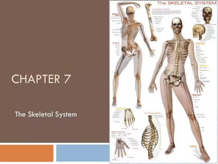
Chapter 7 The Skeletal System
Introduction • Individual bones are the organs of the skeleton system • Bone contains very active tissues
Bone Structure • Bone structure reflects its function • Parts of a long bone • Epiphyses: • Located at each end • Covered by articular cartilage • Articulates (forms joint) with other bones • Diaphysis: • Shaft of bone
Bone Structure Cont. • Parts of a long bone cont. • Periosteum • Covers bone • Mostly found on diaphysis • Compact Bone • Provides strength and resistance to bending • Spongy Bone • Provides strength where needed and reduces weight of bone • Medulllary Cavity • In diaphysis, it’s the area filled with marrow
Microscopic Structure • Osteocytes • Mature bone cells • Lacunae • Bony chambers • Processes • Bony projections that provide a site for ligament and tendon attachment
Microscopic Structure Cont. • Osteons • Found in compact bone • Are cemented together to form cylinder shaped units (Haversian System)
Microscopic Structure Cont. • Osteonic Canals (Haversian Canals) • Contain blood vessels that nourish the cells of the osteons • Spongy bone is nourished by diffusion from the surface of the thin bony plates.
Bone Development and Growth • Intramembranous Bones • Develop from layers of connective tissues • Osteoblasts within the membranous layers form bone tissue
Bone Growth and Development Cont. • Endochondral Bones • Developed first as hyaline cartilage to be later replaced by bone tissue • Primary Ossification (creation of new bone tissue) center appears in the diaphysis, Secondary Oss. In epiphysis.
Bone Growth and Development Cont. • Epiphyseal Disk • Division between primary and secondary ossification centers • Responsible for lengthening bone • Bone continues to lengthen until disks ossify. • Growth in thickness is due to intramembranous ossification beneath periosteum • If Epi. Disk is damaged before ossified, it can cause premature growth ending.
Bone Function • 5 Functions • 1. Support • 2. Protection • Bones shape and form body structures • Bones support and protect softer, underlying tissues
Bone Function Cont. • 3. Body Movement • Bones and muscles function together as levers • A lever consists of a rod, pivot (fulcrum), a movable weight, and a force that supplies energy.
Bone Function Cont. • 4. Blood Cell Formation • Hematopoiesis (development of blood cells) • At different ages, hematopoiesis occurs in the yolk sac, liver and spleen, and red bone marrow. • Red Bone marrow produces red blood cells (RBC’s), white blood cells (WBC’s) and blood platelets • Yellow Marrow stores fat
Bone Function Cont. • 5. Storage of Inorganic Salts • Intercellular material of bone tissue contains large quantities of calcium phosphate. • When blood calcium is low, osteoclasts break down bone, when blood calcium is high, osteoblasts build bone. • Bone stores small amounts of magnesium, sodium, potassium and carbonate ions.
Skeletal Organization p. 142-149 • Axial Portion • Skull • Hyoid bone • Vertebral column • Thoracic cage • Appendicular Portion • Pectoral girdle • Upper limbs • Pelvic girdle • Lower limbs
Skull • Consists of 22 bones • 8 cranial • 13 facial • 1 mandible
Cranium • Encloses and protects the brain • Some bones contain air filled sinuses • Include • Frontal • Parietal • Occipital • Temporal • Sphenoid • Ethmoid
Facial Skeleton • Provides the basic shape of face and attachments for muscles • Includes: • Maxillary • Palatine • Zygomatic • Lacriminal • Nasal • Vomer • Inferior nasal conchae • mandible
Infantile Skull • Fontanels separate incompletely developed bones • Proportions of the infantile skull are different from those of an adult skull.
Table 7.2 Skeletal Structure Terms • Condyle • Definition: A rounded process that usually articulates with another bone • Example: Occipital condyle of occipital bone • Crest • Defintion: A narrow, ridge-like projection • Example: Iliac crest of ilium • Epicondyle • Defintion: A projection situated above a condyle • Example: Medial epicondyle of humerus • Facet • Definition: A small, nearly flat surface • Example: Rib-facet of thoracic vertebra • Fontanel • Definition: A soft spot in the skull where membranes cover the spaces between bones • ExampleAnterior fontanel between frontal and parietal bones
Table 7.2 Skeletal Structure Terms Cont. • Foramen • Definition: An opening through a bone that usually is a passageway for blood vessels, nerves, or ligaments • Example: Foramen magnum of occipital bone • Fossa • Definition: A relatively deep pit or depression • Example: Olecranonfossaofhumerus • Fovea • Definition: A tiny pit or depression • Example: Fovea capitis of femur • Head • Definition: An enlargement of the end of a bone • Example: Head of humerus • Meatus • Definition: A tube-like passageway within a bone • Example: External auditory meatus of ear
Table 7.2 Skeletal Structure Terms Cont. • Process • Definition: A prominent projection on a bone • Example: Mastoid process of temporal bone • Sinus • Definition: A cavity within a bone • Example: Frontal sinus of frontal bone • Spine • Definition: A thorn-like projection • Example: Spine of scapula • Suture • Definition: An interlocking line of union between bones • Example: Lambdoidal suture between occipital and parietal bones • Trochanter • Definition: A relatively large process • Example: Greater trochanter of femur
Table 7.2 Skeletal Structure Terms Cont. • Tubercle • Definition: A small, knob-like process • Example: Greater tubercle of humerus • Tuberosity • Definition: A knob-like process usually larger than a tubercle • Example: Radial Tuberosity of radius
Vertebral Column p. 149-155 • Extends from skull to the pelvis • Protects the spinal cord • Composed of vertebrae • Separated by intervertebral disks • Has four curvatures
Typical Vertebra • Consists of: • A body • A bony arch (surrounds spinal cord) • Contains notches on upper and lower surfaces that provide intervertebral foramina through which spinal nerves pass.
Cervical Vertebrae (7) • Atlas • 1st vertebra-supports and balances head • Axis • 2nd vertebra-contains dens that provide intervertebral foramina through which spinal nerves pass • Contains Transverse Process which bears the transverse foramina
Thoracic Vertebrae (12) • Larger than cervical • Contains facets on the sides that articulate with the ribs
Lumbar Vertebrae (5) • Vertebral Bodies are large and strong • Support more body weight than other vertebrae
Saccrum • Is a triangular structure formed of five fused vertebrae • Vertebral foramina form the sacral canal
Coccyx • Composed of four fused vertebrae • Forms the lowest part of vertebral column • Your TAILBONE!
Thoracic Cage • Includes: • Ribs • Thoracic vertebra • Sternum • Costal cartilages • Supports the shoulder girdle and arms • Protects visceral organs • Functions in breathing
Ribs • 12 Pairs are attached to the thoracic vertebrae • True Ribs join sternum directly by costal cartilages • False Ribs join indirectly or not at all • Typical rib has a shaft, head, and tubercles that articulate with the vertebrae.
Sternum • Consists of three parts: • Manubrium • Body • Xiphoid process • Articulates with clavicles
Pectoral Girdle P. 155 • Composed of: • Two clavicles • Two scapula • Forms an incomplete ring that supports the upper limbs and provides attachment for muscles
Clavicles • Rod-like bones located between the manubrium and scapulae • Hold the shoulders in place and provide attachment for muscles
Scapula • Broad, triangular bones • Articulate with the humerus of each upper limb and provide attachment for muscles
Upper Limbs • Provide framework and attachment for muscles • Function in levers that move the limb and its parts • Contains: • Humerus • Radius • Ulna • Hand
Humerus • Extends from the scapula to the elbow • Articulates with the radius and ulna at elbow
Radius • Located on the thumb side of the forearm between the elbow and wrist • Articulates with humerus, ulna and wrist
Ulna • Longer than radius • Overlaps the humerusposteriorly • Articulates with the radius laterally and with a disk of fibrocartilage inferiorly
Carpals (8) • Consists of 8 bones: • Lunate • Hamate • Triquetrum • Pisiform • Scaphoid • Capitate • Trapezoid • Trapezium • Articulates with the radius and fibrocartilaginous disk on ulnar side
Metacarpals (5) • Consists of 5 bones: • Metacarpals 1-5 • Cylindrical, rounded distal ends that form the knuckles • Articulate proximally with carpals and distaly with phalanges
Phalanges (14) • Finger Bones • Each finger has three phalanges (digits) • A proximal, a middle and a distal phalanx (digit) • The Thumb has two-it lacks a middle phalanx