Download
1 / 24
240 likes | 542 Views
The blueprint of life; from DNA to Protein. Bio 261 Medgar Evers College, CUNY Prof. Santos. DNA, the substance of inheritance Is the most celebrated molecule of our time Hereditary information Is encoded in the chemical language of DNA and reproduced in all the cells of your body

E N D
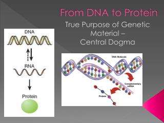
The blueprint of life; from DNA to Protein Bio 261 Medgar Evers College, CUNY Prof. Santos
DNA, the substance of inheritance • Is the most celebrated molecule of our time • Hereditary information • Is encoded in the chemical language of DNA and reproduced in all the cells of your body • It is the DNA program • That directs the development of many different types of traits
G C A T T A 1 nm C G 3.4 nm C G A T G C T A T A A T T A G C 0.34 nm A T Figure 16.7a, c (a) Key features of DNA structure (c) Space-filling model
5 end O OH Hydrogen bond P 3 end –O O OH O A T O CH2 O O P O –O O– O P O H2C O O G C O O CH2 O P O O O –O O– O– O– O P P P O O O H2C O O O O C G O O CH2 O P –O O H2C A T O O CH2 OH 3 end (b) Partial chemical structure 5 end Figure 16.7b
H N O H CH3 N N N N H Sugar N N O Sugar Adenine (A) Thymine (T) H O N H N N N H N Sugar N N N O H Sugar H Figure 16.8 Cytosine (C) Guanine (G)
DNA The Components and Structure of DNA DNA is made up of nucleotides. A nucleotide is a monomer of nucleic acids made up of: • Deoxyribose – 5-carbon Sugar • Phosphate Group • Nitrogenous Base
There are four kinds of bases in in DNA: adenine guanine cytosine thymine
Chargaff’s rule Chargaff's Rules Erwin Chargaff discovered that: • The percentages of guanine [G] and cytosine [C] bases are almost equal in any sample of DNA. • The percentages of adenine [A] and thymine [T] bases are almost equal in any sample of DNA.
There are 2 hydrogen bonds between adenine and thymine and three hydrogen bonds between cytosine and guanine.
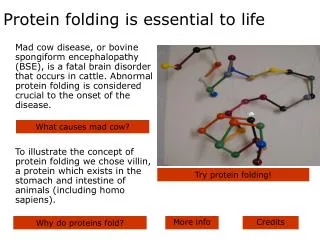
Protein synthesis 3 steps 1- transcription 2- RNA processing 3- translation
Transcription • Copying the genetic code directly from DNA. • We make a single strand of messenger RNA. • We begin initiation by unwinding the double stranded DNA and copying only one of the strands. The strand that is copied is called the sense strand. It serves as a template for the production of messenger RNA.
Transcription begins when an enzyme called RNA polymerase binds to a special region of the DNA called promoter sequence. Unlike DNA polymerase, RNA polymerase doesn’t need a primer. • RNA polymerase brings free floating RNA nucleotides to the sense strand.
Guanine and cytosine pair up. • But, there is no thymine in RNA. Another base called Uracil pairs up with adenine. • The messenger RNA strand will continue to elongate until it reaches a termination point.
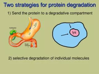
The coding regions are called exons and the non-coding regions are called introns. • The introns are removed by an enzyme-RNA complex known as the spliceosome. • A tail of adenine bases is added to the 3 prime end and a modified guanine nucleotide is added to the 5 prime end.
Once the messenger RNA has been processed, it is ready to leave the nucleus and bind to a ribosome. • The mature messenger RNA carries the message from DNA in the forms of codons. • A codon is a group of 3 bases that correspond to one of the 20 amino acids.
There are 64 possible codons and only 20 amino acids. There is redundancy with some of the amino acids! • The initial codon is AUG or methionine and there are three stop codons, UAA, UGA and UAG.
Second mRNA base U C A G U UAU UUU UCU UGU Cys Tyr Phe UAC UUC UCC UGC C U Ser UUA UCA UAA Stop Stop UGA A Leu UAG UUG UCG Stop Trp UGG G CUU CCU U CAU CGU His CUC CCC CAC CGC C C Arg Pro Leu CUA CCA CAA CGA A Gln CUG CCG CAG CGG G Third mRNA base (3 end) First mRNA base (5 end) U AUU ACU AAU AGU Asn Ser C lle AUC ACC AAC AGC A Thr A AUA ACA AAA AGA Lys Arg Met or start G AUG ACG AAG AGG U GUU GCU GAU GGU Asp C GUC GCC GAC GGC G Ala Gly Val GUA GCA GAA GGA A Glu Figure 17.5 GUG GCG GAG GGG G
Translation • The messenger RNA attaches to the ribosome and the message in the form of codons is “translated” and the appropriate amino acid is put in place. • A molecule of RNA called transfer RNA brings along the amino acid. It resembles a four leaf clover. • On the top is the amino acid and on the bottom is a sequence known as the anti codon.
3 A Amino acid attachment site C C 5 A C G C G C G U G U A A U U A U C G * G U A C A C A * A U C C * G * U G U G G * G A C C G * C A G * U G * * G A G C Hydrogen bonds G C U A G * A * A C * U A G A Anticodon
The anticodon pairs up with the codon. This allows the amino acids to put placed in the correct sequence or order.