Download
1 / 36
370 likes | 425 Views
Explore the organization, structure, and functions of the nervous system, including reflex arcs, neural pathways, and neurotransmitter activity. Learn about the role of position receptors and the vestibular apparatus in movement control.

E N D
Chapter 7:The Nervous System EXERCISE PHYSIOLOGY Theory and Application to Fitness and Performance, 6th edition Scott K. Powers & Edward T. Howley
Objectives • Discuss the general organization of the nervous system • Describe the structure & function of a nerve • Draw and label the pathways involved in a withdraw reflex • Define depolarization, action potential, and repolarization • Discuss the role of position receptors in the control of movement
Objectives • Describe the role of vestibular apparatus in maintaining equilibrium • Discuss the brain centers involved in voluntary control of movement • Describe the structure and function of the autonomic nervous system
General Nervous System Functions 1. Control of the internal environment • Nervous system works with endocrine system 2. Voluntary control of movement 3. Programming spinal cord reflexes 4. Assimilation of experiences necessary for memory and learning
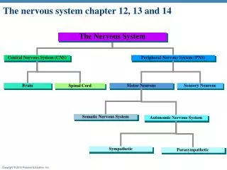
Organization of the Nervous System • Central nervous system (CNS) • Brain and spinal cord • Peripheral nervous system (PNS) • Neurons outside the CNS • Sensory division • Afferent fibers transmit impulses from receptors to CNS • Motor division • Efferent fibers transmit impulses from CNS to effector organs
Divisions of the Nervous System Fig 7.1
Relationship Between PNS and CNS Fig 7.2
Structure of a Neuron • Cell body • Dendrites: Conduct impulses toward cell body • Axon • Carries electrical impulse away from cell body • May be covered by Schwann cells • Forms discontinuous myelin sheath along length of axon • Synapse: Contact points between axon of one neuron and dendrite of another neuron
Structure of a Neuron Fig 7.3
Synaptic Transmission Fig 7.4
Electrical Activity in Neurons • Neurons are “Excitable Tissue” • Irritability: ability to respond to a stimulus and convert it to a neural impulse • Conductivity: transmission of the impulse along the axon
Electrical Activity in Neurons • Resting membrane potential • At rest, the neurons are negatively charged • Determined by concentrations of ions (Na+, K+, Cl-) across membrane • Action potential • Occurs when depolarization reaches threshold • Permeability of the membrane changes, allowing Na+ into the cell, making the interior positively charged • Repolarization • Change in membrane permeability, restoring resting membrane potential
An Action Potential Fig 7.5
Depolarization Fig 7.6a
Repolarization Fig 7.6b
Neurotransmitters and Synaptic Transmission • Neurons communicate across synapses using neurotransmitters • Released from presynaptic membrane • Binds to receptor on post synaptic membrane
Neurotransmitters and Synaptic Transmission • Excitatory postsynaptic potentials (EPSP) • Causes depolarization which may or may not reach threshold • Temporal summation: summing several EPSPs from one presynaptic neuron • Spatial summation: summing from several different presynaptic neurons • Inhibitory postsynpatic potentials (IPSP) • Causes hyperpolarization
Sensory Information • Proprioceptors • Proprioception: ability to determine position of joint • Kinesthesia: sensation of joint motion or acceleration • Muscle Chemoreceptors • Sensitive to changes in the chemical environment surrounding a muscle
Proprioceptors • Provide CNS with information about body position and joint angle • Free nerve endings – touch & pressure • Golgi-type receptors – in ligaments & joints • Pacinian corpuscles – in tissues around joints • Strongly stimulated then adapt
Muscle Chemoreceptors • Provide CNS with information regarding the metabolic rate of muscular activity • Hydrogen ion concentration • Carbon dioxide (CO2) • Potassium (K+)
Reflexes • Rapid, unconscious means of reacting to stimuli • Order of events: • Sensory nerve sends impulse to spinal column • Interneurons activate motor neurons • Motor neurons control movement of muscles • Reciprocal inhibition • EPSPs to muscles to withdraw from stimulus • IPSPs to antagonistic muscles Fig 7.8
Somatic Motor Function • Somatic motor neurons of PNS • Responsible for carrying neural messages from spinal cord to skeletal muscles • Motor unit • Motor neuron and all the muscle fibers it innervates • Innervation ratio • Number of muscle fibers per motor neuron
Illustration of a Motor Unit Fig 7.9
Vestibular Apparatus and Equilibrium • Located in the inner ear (Semi-circular canals) • Responsible for maintaining general equilibrium and balance • Sensitive to changes in linear and angular acceleration
Motor Control Functions of the Brain • Brain stem: responsible for • Many metabolic functions • Cardiorespiratory control • Major structures • Medulla • Pons • Midbrain • Reticular formation – a series of complex neurons scattered throughout the brain stem
Motor Control Functions of the Brain • Cerebrum • Cerebral cortex • Organization of complex movement • Storage of learned experiences • Reception of sensory information • Motor cortex • Most concerned with voluntary movement • Cerebellum - Monitors complex movement
Motor Functions of the Spinal Cord • Withdrawal reflex • Contains groups of neurons capable of controlling certain aspects of motor activity • Spinal tuning • Voluntary movement is translated into appropriate muscle action
Control of Motor Function • Subcortical and cortical motivation areas • Sends a “rough draft” of the movement • Cerebellum and basal ganglia • Coverts “rough draft” into movement plan • Cerebellum: fast movements • Basal ganglia: slow, deliberate movements • Motor cortex through Thalamus • Forwards message sent down spinal neurons for “Spinal tuning” and onto muscles • Feedback from muscle receptors and proprioceptors allows fine-tuning of motor program
Structures and Processes Leading to Voluntary Movement Fig 7.12
Autonomic Nervous System • Responsible for maintaining internal environment • Effector organs not under voluntary control • Smooth muscle, cardiac muscle, glands • Sympathetic division • Releases norepinephrine (NE) • Excites an effector organ • Parasympathetic division • Releases acetylcholine (ACh) • Inhibits effector organ
Exercise Enhance Brain Health • A recent five-year study in humans has concluded that exercise improves brain function and reduces the risk of cognitive impairment associated with aging • It is clear that regular exercise can protect the brain against disease (e.g. Alzheimer’s) and certain types of brain injury (e.g. stroke)