Download
1 / 55
550 likes | 573 Views
Learn about P, QRS, T waves, leads nomenclature, ECG interpretation, rate calculations, axis deviations, and rhythm analysis in this primer on ECG fundamentals.

E N D
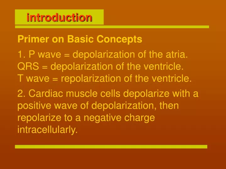
introduction Primer on Basic Concepts 1. P wave = depolarization of the atria.QRS = depolarization of the ventricle.T wave = repolarization of the ventricle. 2. Cardiac muscle cells depolarize with a positive wave of depolarization, then repolarize to a negative charge intracellularly.
Height and depth of wave is amplitude measured in millimeters one small squares equal to 1 milimeter and two large squares equal to 1 cm which is equal to 1 mv 2 large squares =1 cm = 1 mv . Horizontal axis is time..04 seconds for 1 mm (1 small box)..2 seconds for 1 large box = 5 small boxes = 5 x .04 seconds.
7. Lead nomenclature.Limb Leads: I, II, III, aVR, aVF, aVLChest Leads: V1 - V6 Rhythm Strip: Located on the bottom of the ECG printout. Selected to give the best relationship of the P wave to the QRS. Figure 3: A normal ECG and rhythm strip.
I. Rate • II.. AXIS. • III. Rhythm + CODUCTION • IV. Hypertrophy • V. Infarct 8. ECG interpretation: look at five areas, in order, on each ECG.
I. Rate Rate is cycles or beats per minute. Normal rate for the SA node 60-100. [ <60 bradycardia >100 tachycardia ] SA node is the usual pacemaker, other potential pacemakers (if SA node fails) are atrial pacemakers with inherent rates of 60-80, AV node (rate 40-60), or ventricular pacer (rate 20-40). In certain pathologic conditions ectopic (out of place) pacemakers can go much faster at rates 150-250 cycles/minute. There are three methods of calculating rate: 1. Most Common Method:(Most rates can be calculated this way). Find an R wave on a heavy line (large box) count off "300, 150, 100, 75, 60, 50" for each large box you land on until you reach the next R wave. Estimate the rate if the second R wave doesn't fall on a heavy black line. Rate calculation: Memorize the number sequence, 300, 150, 100, 75, 60, 50
I. Rate Figure 4: common method
I. Rate 2. Mathematical method:Use this method if there is a regular bradycardia, i.e. - rate < 50. If the distance between the two R waves is too long to use the common method, use the approach: 300/[# large boxes between two R waves]. Figure 5: Count number of large boxes between first and second R waves =7.5. [ 300/7.5 large boxes = rate 40 ].
I. Rate 3. Six-second method:Count off 30 large boxes = 6 seconds (remember 1 large box = 0.2 seconds, so 30 large boxes = 6 seconds). Then, count the number of R-R intervals in six seconds and multiply by 10. This is the number of beats per minute. This is most useful if you have an irregular rhythm (like atrial fibrillation) when you want to know an average rate. Figure 6: Count 30 large boxes, starting from the first R wave. There are 8 R-R intervals within 30 boxes. Multiply 8 x 10 = Rate 80.
I. Rate The rate is about 90. The second R wave in the rhythm strip lands on a heavy line. Count "300-150-100-75" over (four heavy lines) to the next R wave to determine the rate. You'll have to estimate between 75 and 100 for this example.
I. Rate The rate is about 40. Use the bradycardia method where 300 divided by the number of large boxes between R waves is the rate. In this case, there are 7.5 large boxes between R waves. 300 divided by 7.5 equals a rate of 40.
I. Rate Rate about 70-80. Use the six second strip method. There are 30 large boxes in six seconds. Multiply the number of R-R intervals in six seconds by ten to get the rate.
. Axis Lead I Lead aVF 1. Normal axis (0 to +90 degrees) Positive Positive 2. Left axis deviation (-30 to -90) Also check lead II. To be true left axis deviation, it should also be down in lead II. If the QRS is upright in II, the axis is still normal (0 to -30). Positive Negative 3. Right axis deviation (+90 to +180) Negative Positive 4. Indeterminate axis (-90 to -180) Negative Negative
III. Axis Figure 29: Normal axis
III. Axis Figure 30: Left axis deviation
III. Axis Figure 31: Right axis deviation
III. Axis • The bottom line is, if the axis is shifted out of the normal quadrant, evaluate the reasons for this. • Differential Diagnosis • Left axis deviation LVH, left anterior fasicular block, inferior wall MI • Right axis deviation RVH, left posterior fascicular block, lateral wall MI
III. Axis Quiz Normal Axis. Positive QRS in Lead I and aVF
III. Axis Quiz Left axis deviation. Positive QRS in lead I, but negative QRS in leads II and aVF
III. Axis Quiz Right axis deviation. Positive QRS in lead aVF, but negative QRS in lead I.
II. Rhythm We will focus on the basic "core" of rhythms and measured "intervals" (PR, QRS, QT). Rhythms are often the most challenging aspect of ECG's. Now for some basics - "arrhythmia" means abnormal rhythm. The normal conduction pathway is: SA node --> AV node --> Bundle of HIS --> Bundle Branches. Arrhythmia can be understood by realizing the existence of ectopic (out of place) foci (pacemakers) and understanding the normal conduction pathway of the heart. Very simply put, if the beat originates in the atria or AV node (supraventricular) the QRS is usually narrow (normal), because it comes from above along the normal pathway. Figure 6a: QRS is narrow (normal)
II. Rhythm (to include intervals) If the beat is ventricular in origin, the QRS is wide and bizarre because it doesn't come down the normal pathway. Figure 6b: QRS is wide. A reasonable way to group arrhythmias is in four general groups. Let us briefly review these four groups, then we will develop some common sense principles for evaluating rhythm (to include intervals).
II. Rhythm Group 1 irregular rhythms (main clue is QRS is not spaced evenly apart anywhere, total irregularity of the beat). A) Sinus arrhythmia. P waves and P-R intervals are all identical because they originate from the sinus node. Sinus rate may vary normally a bit (increase with inspiration, decrease with expiration), but if the rate varies a lot, this term is used. Figure 7: Sinus arrhythmia: P waves are identical
II. Rhythm Group 1 irregular rhythms B) Wandering atrial pacemaker Pacemaker discharges from different atrial locations - the clue here is the P waves are of varying shape and differing PR intervals. PR interval is measured from the beginning of the the Pwave to the beginning of the QRS - if the atrial pacemaker location varies it will take different lengths of time to get to the ventricle - resulting in different PR intervals. If the rate of the wandering atrial pacemaker is >100 it is descriptively called multifocal atrial tachycardia. Figure 8: Multifocal atrial tachycardia
II. Rhythm Group 1 irregular rhythms C) Atrial fibrillation. You will frequently see this arrhythmia. There are no P waves, only irregular or wavy baseline. The QRSs are irregularly spaced, therefore it is included under irregular rhythms. Figure 9: Atrial fibrillation
II. Rhythm Group 3 Rapid ectopic (out of place) rhythms Figure 16: Paroxysmal supraventricular tachycardia: note accelerated rate and narrow QRS complexes
II. Rhythm Group 3 Rapid ectopic (out of place) rhythms Figure 17: Ventricular tachycardia: note fast rate and wide bizarre QRS
II. Rhythm Group 3 Rapid ectopic (out of place) rhythms Figure 18: Ventricular fibrillation: erratic and wavy baseline
CONDUCTION (occur in three (3) degrees, like skin burns; third degree is the worst) 1st degree PR interval > 0.2 seconds (1 large box), each P is followed by a QRS. PR interval is measured from the beginning of the P wave to the beginning of the QRS. Figure 19: The PR interval is approximately 0.28 seconds
II. CONDUCTION 2nd degree block - type 1 Also called "Wenkebach". PR interval gets progressively longer each beat until finally a QRS is "dropped" (missing). Figure 20: Note the increasing PR interval before the QRS is dropped, then the cycle is repeated
CONDUCTION 2nd degree block - type 2 Also called "Mobitz II". Look out! A more serious conduction problem than Type 1. PR intervals are constant and a QRS is "dropped" intermittently. Figure 21: Note the dropped QRS after the second and sixth P wave in lead II (the rhythm strip)
II. CONDUCTION 3rd degree block The atrial rate is independent of the ventricular rate (P wave and QRS march out separately. The clue here is no relationship at all of the P-R intervals). The P-R interval is constantly changing, the QRS is usually wide and bizarre because it is ventricular origin. Figure 22: Note the P waves and QRS waves are independent of each other
CONDUCTION An interval is a portion of the baseline and at least one wave. We measure an interval on the horizontal axis in seconds. The PR, QRS, and QT are the intervals which should be routinely scanned on each ECG. For measuring intervals, look at the widest form in any lead. 1. PR interval (beginning of P wave to the beginning of the next QRS). Normally, < .2 seconds or one large box. If it is > .2 seconds, it is a first degree block. (Note: this concept was introduced under blocks) Figure 24: Note the prolonged PR interval (.28 seconds), especially at the second beat Figure 23: Intervals
II. CODUCTION 2. QRS interval (beginning of Q to the end of the S wave) should be < .12 seconds (< 3 small boxes). If QRS is > .12, check for bundle branch block. A QRS > .12 and RR (2 peaks or R waves in QRS) occurring in the right chest leads (V1-V2) indicates a right bundle branch block. Figure 25: RBBB
CONDUCTION If QRS is > .12 and RR occurs in the left chest leads (V5-V6), this indicates a left bundle branch block. Figure 26: LBBB Very important: For left bundle branch block, the ECG is often unreliable for identifying infarct. Infarct is a concept explained later in this tutorial, but don't forget that with left bundle branch block, one cannot rely on the ECG for diagnosis of infarct.
II. CONDUCTION 3. QT interval (beginning of QRS to end of T wave) should be less than half of the preceding RR interval - this varies with the rate. For normal rates, QT < .4 seconds (2 large boxes). "QT prolongation" (too long) can lead to a refractory form of ventricular tachycardia called torsades de pointes. Figure 27: The QT interval is greater than half the preceding RR interval. Look at lead I
II. Rhythm period Rhythm Guidelines 1. Check the bottom of the rhythm strip for regularity, i.e. - is it completely regular, mostly regular with a few extra beats, or totally irregular? 2. Check for a P wave before each QRS, QRS after each P. 3. Check RR interval (for AV blocks) and QRS interval (for bundle branch blocks). Check for prolonged QT. 4. Continue to recognize "patterns" such as atrial fibrillation, PVCs, PACs, escape beats, ventricular tachycardia, paroxysmal atrial tachycardia, AV blocks and bundle branch blocks.
II. Rhythm & CONDUCTION Quiz Question 1 Match the tracings with the correct interpretation: A. First Degree BlockB. Sinus ArrhytmiaC. Third Degree BlockD. Atrial Fibrillation
II. Rhythm & Intervals Quiz Question 1 answer: The correct matches are C. Third Degree BlockThe atrial rate is independent of the ventric. rate so the P waves are independent of the QRS's. The P waves are regular and can be marched out with calipers. The same can be done with the QRS complexes. D. Atrial FibrillationNo P wave, irregular QRS with a wavy baseline. A. First Degree BlockPR interval is greater than .2 seconds (one large box). Note that there is a P wave before each QRS and a QRS after each P wave.
II. Rhythm & Intervals Quiz Question 2 Match the tracings with the correct interpretation: A. Second Degree Block Type 1C. Second Degree Block Type 2D. Paroxysmal Supraventricular Tachycardia
II. Rhythm & Intervals Quiz Question 2: The correct matches are C. Second Degree Block Type 2PR intervals are the same, but every other beat is dropped. D. Paroxysmal Supraventricular TachycardiaRegular narrow QRS. Often it's hard to determine whether it's atrial or junctional as it is going too fast to see P waves. Clinically it doesn't matter since they are treated the same.
II. Rhythm & Intervals Quiz Question 3 Match the tracings with the correct interpretation: A. Ventricular TachycardiaB. Second Degree Block Type 1 (Wenckebach)C. Junctional RhythmD. Wandering Atrial Pacemaker
II. Rhythm & Intervals Quiz Question 3 The correct matches are: D. Wandering Atrial Pacemaker(It's called Multi focal Atrial Tachycardia if the rate is >100). Strict definition is 3 different P wave morphologies and 3 different PR intervals. A. Ventricular TachycardiaFast, wide, regular, bizarre QRS. B. Second Degree Block Type 1 (Wenckebach)The PR gets progressively longer until a beat is dropped.