Download
1 / 74
750 likes | 773 Views
This lecture by Dr. Avraham Samson delves into the structural bioinformatics of proteins and nucleic acids, exploring the common building blocks of all life forms and the forces influencing their structure. Topics covered include gene transfer, atomic composition, chemical forces, protein levels, and molecular interactions.

E N D
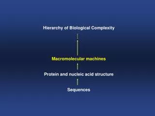
Fundamentals of protein and nucleic acid structure Lecture 2 Structural Bioinformatics Dr. Avraham Samson 81-871
Tree of Life All known life forms use the same building blocks suggesting there was a common ancestor
The tree of life is not that simple! Gene transfer across kingdoms continually occurs (cyanobacteria became chlorophylls, proteobacteria mitochondria, viruses)
29 atoms of life Most common elements:H, O, C, N, P, S (97% of organismweight) Most common ions: Ca2+, K+, Na+, Mg2+, Cl-
Forces affecting structure: • H-bonding • Van der Waals • Electrostatics • Hydrophobicity • Disulfide Bridges d 2.6 Å < d < 3.1Å 150° < θ < 180°
Forces affecting structure: • H-bonding • Van der Waals • Electrostatics • Hydrophobicity • Disulfide Bridges Repulsion דחייה Attraction משיכה d 3 Å < d < 4Å
Forces affecting structure: • H-bonding • Van der Waals • Electrostatics • Hydrophobicity • Disulfide Bridges d d = 2.8 Å “IONIC BOND” יוניקשר “SALT BRIDGE” מלחגשר E = Energy k = constant D = Dielectric constant (vacuum = 1; H2O = 80) q1 & q2 = electronic charges (Coulombs) r = distance (Å) Coulomb’s law
Forces affecting structure: • H-bonding • Van der Waals • Electrostatics • Hydrophobicity • Disulfide Bridges
Forces affecting structure: • H-bonding • Van der Waals • Electrostatics • Hydrophobicity • Disulfide Bridges Other names: cystine bridge disulfide bridge Hair contains lots of disulfide bonds which are broken and reformed by heat
Levels of protein structure Primary: Amino acid sequence Secondary: Local fold pattern of small subsequence Tertiary: Fold of entire protein chain Quaternary: Complex of multiple chains
Primary (1o)structure The amino acid sequence is the primary structure
Primary (1o)structure 20 amino acids
pKa = pH of 50% dissociation
Amino acid nomenclature Primary (1o)structure
Primary (1o)structure • Amino acids polymerize through peptide bonds to form polypeptides
Primary (1o)structure • N-terminal is the start of a polypeptide chain • Amino acids are also called residues
side chains צדדישייר שלד backbone Primary (1o)structure
Primary (1o)structure • Post translational modification are important because they can change the function of proteins (i.e. phosphorylation, acetylation, hydroxylation, carbohydrate and lipid modifications) N-terminal acetylation hydroxyproline O-phosphotyrosine γ-carboxyglutamate
Amino acids chirality Enantiomers –mirror images - תמונת ראי- אננטיומרים chiral center Cכיראלימרכז Dextro-Laevus in Latin
Most proteins: only L amino acids C chiral center is L configuration • איך לקבועקונפיגורציה L וD- • סדר את האטומים לפי עדיפותעלפיהכללים הבאים • כלל .1 אטום עם מספר אטומי יותר גבוה בעל סדר עדיפות יותר גבוה. • (I > Cl > O > N> C > H) • כלל .2 אם האטומים זהים, העדיפותעלפי האטומים המתמירים • (C(CH3)3 > CH(CH3)2 > CH2CH3 > CH3) • שים את האטום עם סדר העדיפות הכי נמוך מאחור. • קבע את כיווןהחץ , מסדר עדיפות הכי גבוה לככוון הכי נמוך. אם החץ: • עםכיוון השעוןקונפיגורציה D • נגד כיוון השעון קונפיגורציה L רמז יותר קל בשביל לזכור :) CORNתירס (
Torsion angles w, φ, and ψ • Unlike w, the two backbone dihedral angles φ and ψ are free to rotate • This rotation freedom allows protein folding Dihedral angles: -180o <φ < +180o -180o < ψ < +180o w is 0o or 180o w
Peptide bond is planar Cα, C, O, N, H, Cα all lie in the same plane
Torsion angle (w) is usually trans Steric hindrance Question: What other residues can be cis?
Except for X-Pro bond in which cis is preferred Steric hindrance Steric hindrance Steric hindrance allows both cis and trans (4:1 ratio)
Ramachandran Diagrams • Steric hindrance dictates torsion angle preference • Ramachandran plot show preferred regions of φ and ψ dihedral angles Preferred regions
Secondary (2o)structure Helices • a-helix • 310-helix • p-helix b Sheets • Antiparallel • Parallel Turns • b,g,a,and p Unstructured • α-helix is the most common • 3.6 residues per turn (number of residues in one full rotation of 360°) • 5.4 Å pitch (translation along axis for one full rotation of 360°) Hydrogen bond: i→i+4
Secondary (2o)structure Helices • a-helix • 310-helix • p-helix b Sheets • Antiparallel • Parallel Turns • b,g,a,and p Unstructured 310-helices are rare in proteins: 3.1 residues per turn (number of residues in one full rotation of 360°) 6 Å pitch (translation along axis for one full rotation of 360°) Hydrogen bond: i→i+3
Secondary (2o)structure Helices • a-helix • 310-helix • p-helix b Sheets • Antiparallel • Parallel Turns • b,g,a,and p Unstructured π-helices are rare in proteins 4.3 residues per turn (number of residues in one full rotation of 360°) 6 Å pitch (translation along axis for one full rotation of 360°) Hydrogen bond: i→i+5
Hydrogen bonding in helices • CO of residue (n) forms an h-bond with NH of residue: • (n+4) → α-helix • (n+3) → 310-helix • (n+5) → π-helix
Secondary (2o)structure Helices • a-helix • 310-helix • p-helix b Sheets • Antiparallel • Parallel Turns • b,g,a,and p Unstructured • In antiparallel b-sheets • Adjacent β-strands run in opposite directions • Hydrogen bonds (dashed lines) between NH and CO stabilize the structure • The side chains (in green) are above and below the sheet
Secondary (2o)structure Helices • a-helix • 310-helix • p-helix b Sheets • Antiparallel • Parallel Turns • b,g,a,and p Unstructured • In parallel b-sheets • Adjacent β-strands run in same direction • Hydrogen bonds (dashed lines) between NH and CO stabilize the structure • The side chains (in green) are above and below the sheet
Ribbon diagram of β sheet • In addition to being purely parallel or antiparallel, β sheets can be mixed, with strands running in both parallel and antiparallel directions • Arrow pointing to C-terminal end
Secondary (2o)structure CO of residue i forms h-bonds with NH of either residue i+2, i+3, i+5, or i+4 β-turn i→i+3 H-bond (most common) γ-turn: i→i+2 H-bond α-turn i→i+4 H-bond π-turn i→i+5 H-bond Helices • a-helix • 310-helix • p-helix b Sheets • Antiparallel • Parallel Turns • b,g,a,and p Unstructured
Secondary (2o)structure Helices • a-helix • 310-helix • p-helix b Sheets • Antiparallel • Parallel Turns • b,g,a,and p Unstructured
Tertiary (3o)structure • Proteins fold into compact tertiary structures with hydrophobic cores. (spacefill representation) Structural classification: http://www.cathdb.info/cgi-bin/cath/GotoCath.pl?link=class.html
Tertiary (3o)structure • It is a bit difficult to see and understand anything here (sticks representation)
Tertiary (3o)structure • To simplify representation, secondary structure diagrams are used
Quaternary (4o)structure Dimer of identical subunits (Cro protein of bacteriophage lambda)
Quaternary structure • Coat of rhinovirus • (common cold) • 60 copies of 4 • different subunits, • 3 outside, • red, blue, green
Protein structure databases • Primary: UniProthttp://www.uniprot.org/ • Secondary: DSSP http://rcsb.org • Tertiary: PDB http://rcsb.org • Quaternary: PQS http://www.ebi.ac.uk/pdbe/pqs/
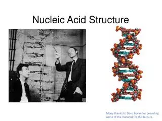
DNA and RNA Structure • NOTE: • Components • Sugar • Base • Phosphate • 5’ to 3’ direction • T->U in RNA • RNA - extra –OH at 2’ of pentose sugar • DNA - deoxyribose • Numbering • Single vs double strands • DNA more stable Voet, Donald and Judith G. Biochemistry. John Wiley & Sons, 1990, p. 792.
NOTE: • Pyrimadines and Purines • T->U in RNA • Names • Numbering • Bonding character • Position of hydrogen • Tautomers Purines The 5 Basesof DNA and RNA Pyrimadines