Download
1 / 54
560 likes | 685 Views
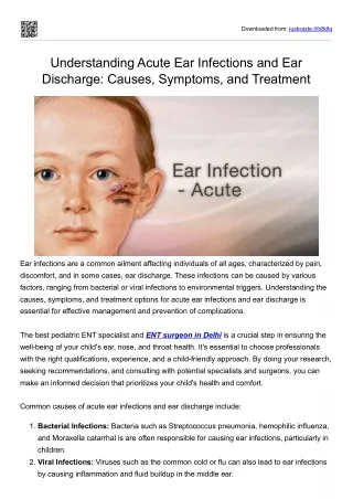
Understand infections of the external ear, including Otitis Externa, Furunculosis, Otomycosis, Granular Myringitis, Bullous Myringitis, and Necrotizing External Otitis. Learn about symptoms, signs, and treatment options for each condition.

E N D
Infections of the External Ear Michael Underbrink, MD Jeffrey Vrabec, MD March 21, 2001
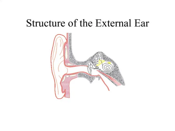
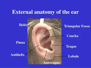
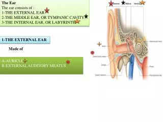
Anatomy and Physiology • Consists of the auricle and EAM • Skin-lined apparatus • Approximately 2.5 cm in length • Ends at tympanic membrane
Anatomy and Physiology • Auricle is mostly skin-lined cartilage • External auditory meatus • Cartilage: ~40% • Bony: ~60% • S-shaped • Narrowest portion at bony-cartilage junction
Anatomy and Physiology • EAC is related to various contiguous structures • Tympanic membrane • Mastoid • Glenoid fossa • Cranial fossa • Infratemporal fossa
Anatomy and Physiology • Innervation: cranial nerves V, VII, IX, X, and greater auricular nerve • Arterial supply: superficial temporal, posterior and deep auricular branches • Venous drainage: superficial temporal and posterior auricular veins • Lymphatics
Anatomy and Physiology • Squamous epithelium • Bony skin – 0.2mm • Cartilage skin • 0.5 to 1.0 mm • Apopilosebaceous unit
Otitis Externa • Bacterial infection of external auditory canal • Categorized by time course • Acute • Subacute • Chronic
Acute Otitis Externa (AOE) • “swimmer’s ear” • Preinflammatory stage • Acute inflammatory stage • Mild • Moderate • Severe
AOE: Preinflammatory Stage • Edema of stratum corneum and plugging of apopilosebaceous unit • Symptoms: pruritus and sense of fullness • Signs: mild edema • Starts the itch/scratch cycle
AOE: Mild to Moderate Stage • Progressive infection • Symptoms • Pain • Increased pruritus • Signs • Erythema • Increasing edema • Canal debris, discharge
AOE: Severe Stage • Severe pain, worse with ear movement • Signs • Lumen obliteration • Purulent otorrhea • Involvement of periauricular soft tissue
AOE: Treatment • Most common pathogens: P. aeruginosa and S. aureus • Four principles • Frequent canal cleaning • Topical antibiotics • Pain control • Instructions for prevention
Chronic Otitis Externa (COE) • Chronic inflammatory process • Persistent symptoms (> 2 months) • Bacterial, fungal, dermatological etiologies
COE: Symptoms • Unrelenting pruritus • Mild discomfort • Dryness of canal skin
COE: Signs • Asteatosis • Dry, flaky skin • Hypertrophied skin • Mucopurulent otorrhea (occasional)
COE: Treatment • Similar to that of AOE • Topical antibiotics, frequent cleanings • Topical Steroids • Surgical intervention • Failure of medical treatment • Goal is to enlarge and resurface the EAC
Furunculosis • Acute localized infection • Lateral 1/3 of posterosuperior canal • Obstructed apopilosebaceous unit • Pathogen: S. aureus
Furunculosis: Symptoms • Localized pain • Pruritus • Hearing loss (if lesion occludes canal)
Furunculosis: Signs • Edema • Erythema • Tenderness • Occasional fluctuance
Furunculosis: Treatment • Local heat • Analgesics • Oral anti-staphylococcal antibiotics • Incision and drainage reserved for localized abscess • IV antibiotics for soft tissue extension
Otomycosis • Fungal infection of EAC skin • Primary or secondary • Most common organisms: Aspergillus and Candida
Otomycosis: Symptoms • Often indistinguishable from bacterial OE • Pruritus deep within the ear • Dull pain • Hearing loss (obstructive) • Tinnitus
Otomycosis: Signs • Canal erythema • Mild edema • White, gray or black fungal debris
Otomycosis: Treatment • Thorough cleaning and drying of canal • Topical antifungals
Granular Myringitis (GM) • Localized chronic inflammation of pars tensa with granulation tissue • Toynbee described in 1860 • Sequela of primary acute myringitis, previous OE, perforation of TM • Common organisms: Pseudomonas, Proteus
GM: Symptoms • Foul smelling discharge from one ear • Often asymptomatic • Slight irritation or fullness • No hearing loss or significant pain
GM: Signs • TM obscured by pus • “peeping” granulations • No TM perforations
GM: Treatment • Careful and frequent debridement • Topical anti-pseudomonal antibiotics • Occasionally combined with steroids • At least 2 weeks of therapy • May warrant careful destruction of granulation tissue if no response
Bullous Myringitis • Viral infection • Confined to tympanic membrane • Primarily involves younger children
Bullous Myringitis: Symptoms • Sudden onset of severe pain • No fever • No hearing impairment • Bloody otorrhea (significant) if rupture
Bullous Myringitis: Signs • Inflammation limited to TM & nearby canal • Multiple reddened, inflamed blebs • Hemorrhagic vesicles
Bullous Myringitis: Treatment • Self-limiting • Analgesics • Topical antibiotics to prevent secondary infection • Incision of blebs is unnecessary
Necrotizing External Otitis(NEO) • Potentially lethal infection of EAC and surrounding structures • Typically seen in diabetics and immunocompromised patients • Pseudomonas aeruginosa is the usual culprit
NEO: History • Meltzer and Kelemen, 1959 • Chandler, 1968 – credited with naming
NEO: Symptoms • Poorly controlled diabetic with h/o OE • Deep-seated aural pain • Chronic otorrhea • Aural fullness
NEO: Signs • Inflammation and granulation • Purulent secretions • Occluded canal and obscured TM • Cranial nerve involvement
NEO: Imaging • Plain films • Computerized tomography – most used • Technetium-99 – reveals osteomyelitis • Gallium scan – useful for evaluating Rx • Magnetic Resonance Imaging
NEO: Diagnosis • Clinical findings • Laboratory evidence • Imaging • Physician’s suspicion • Cohen and Friedman – criteria from review
NEO: Treatment • Intravenous antibiotics for at least 4 weeks – with serial gallium scans monthly • Local canal debridement until healed • Pain control • Use of topical agents controversial • Hyperbaric oxygen experimental • Surgical debridement for refractory cases
NEO: Mortality • Death rate essentially unchanged despite newer antibiotics (37% to 23%) • Higher with multiple cranial neuropathies (60%) • Recurrence not uncommon (9% to 27%) • May recur up to 12 months after treatment
Perichondritis/Chondritis • Infection of perichondrium/cartilage • Result of trauma to auricle • May be spontaneous (overt diabetes)
Perichondritis: Symptoms • Pain over auricle and deep in canal • Pruritus
Perichondritis: Signs • Tender auricle • Induration • Edema • Advanced cases • Crusting & weeping • Involvement of soft tissues
Relapsing Polychondritis • Episodic and progressive inflammation of cartilages • Autoimmune etiology? • External ear, larynx, trachea, bronchi, and nose may be involved • Involvement of larynx and trachea causes increasing respiratory obstruction
Relapsing Polychondritis • Fever, pain • Swelling, erythema • Anemia, elevated ESR • Treat with oral corticosteroids
Herpes Zoster Oticus • J. Ramsay Hunt described in 1907 • Viral infection caused by varicella zoster • Infection along one or more cranial nerve dermatomes (shingles) • Ramsey Hunt syndrome: herpes zoster of the pinna with otalgia and facial paralysis
Herpes Zoster Oticus: Symptoms • Early: burning pain in one ear, headache, malaise and fever • Late (3 to 7 days): vesicles, facial paralysis
Herpes Zoster Oticus: Treatment • Corneal protection • Oral steroid taper (10 to 14 days) • Antivirals