Download
1 / 17
180 likes | 409 Views
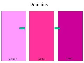
EGF Domains. Emma Howard. EGF Structure. These repeat domains – 30-40 amino acids in length Conserved arrangement of 6 cysteine residues These residues form 3 evenly spaced disulphide bonds. Proteins which have such domains are large >1000a.a., have multiple copies of this domain.

E N D
EGF Domains Emma Howard
EGF Structure • These repeat domains – 30-40 amino acids in length • Conserved arrangement of 6 cysteine residues • These residues form 3 evenly spaced disulphide bonds. • Proteins which have such domains are large >1000a.a., have multiple copies of this domain. • EGF motif found frequently in extra-cellular proteins
EGF Structure • Structurally the EGF domain stabilised by 3 disulfides with disulphide connectivity ababcc. • The domain – 2 β-sheets, major (N terminal) and minor C-terminal)
EGF Evolution • Two types of EGF domain differentiated by location Human (hEGF) half cystine in hairpin of minor sheet. Complement (cEGF) domains half cystine in second strand minor sheet itself. • Postulated to have arisen from a four disuphide ancester, e.g. laminin and integrin.
Functional Differences cEGF hEGF Both types of EGF domain bind calcium. A subset of EGF modules contain a consensus sequence to bind calcium (cbEGF)
Marfans • Autosomal dominant disorder, connective tissue, mutations in fibrilin 1 on chr 15. • Over 500 mutations documented, mostly missense (two thirds). • Mainly in the EGF domains affect calcium binding and alter secondary structure of the protein.
Marfans • Schematic representation of the domain organization of fibrillin-1.
Marfans • Calcium binding EGF motifs, saturate the EGF domains with calcium – rigidity to the protein structure. • Ca binding stabilises fibrilin 1 against proteolytic degradation. • Mutations affecting the calcium consensus sequences – reduce calcium affinity • Mutations in conserved cysteine residues cause domain mis-folding – affect global structure
Haemophillia B (Christmas Disease) • X – linked recessive disorder, Factor IX gene 1 in 50,000 males affected. • Spontaneous bleeding in joints, leading to arthritis and joint deformity. • 2 EGF domains in Factor IX gene , one calcium binding – essential for biological activity, one non-calcium binding which binds with factor VIIIa.
Factor IX • EGF-1 binds calcium at ligands Asp45, Gly48, Gln50, Asp64, and Asp65. • Mutations in this domain, effects saturation of calcium – can’t bind, effecting activation of Gla domain and conformational change of the protein. • EGF domains function as spacers to position the active site of FIX and related coagulation proteases at a suitable distance from the biological membrane for interaction with activators, cofactors, and substrates.
Factor IX • p.P55S and p.P55L cause a milder phenotype • Pro55Ser mutation causes hemophilia primarily due to an impaired ability to activate FX • Pro55Leu defect interferes with the activation of FIX • Marginal changes in the calcium binding, causing aberrant proteolysis – increased degradation of protein.
Hypercholestrolemia • Familial hypercholesterolemia, FH (type II hyperlipoproteinemia) is an autosomal dominant disorder that results from mutations affecting the structure and function of the cell-surface receptor that binds plasma LDLs (low density lipoproteins) removing them from the circulation.
Hypercholestrolemia • Class 2B mutations are the most common • Mutations in the EGF-like domain cause class 2b mutations, associated with defective transport of the protein. • Over 60 mutations in this domain, affect binding of calcium – results in misfolding of the domain. • There is also the possibility that the misfolding of the calcium-binding epidermal growth factor-like pair region is propagated to other regions of the intact receptor, resulting in more global defects.
Functions • Spacer within protein allowing proper conformation • Calcium binding gives rigidity in protein – protects against proteolysis • Involved in protein/protein interactions and interactions between intra-protein domains.
References • Functional analysis of the EGF-like domain mutations Pro55Ser and Pro55Leu, which cause mild hemophilia B. Knobe K.E.; Persson K.E.M.; Sjörin E.1; Villoutreix B.O.2; Stenflo J.3; Ljung R.C.R.Journal of Thrombosis and Haemostasis, Volume 1, Number 4, April 2003 , pp. 782-790(9) • The N-terminal Epidermal Growth Factor-like Domain of Coagulation Factor IX. Kristina E. M. Persson, Bruno O. Villoutreix, Ann-Marie Tha mlitz, Karin E. Knobe and Johan Stenflo. Vol. 277, No. 38, Issue of September 20, pp. 35616–35624, 2002. • Global Defects in the Expression and Function of the Low Density Lipoprotein Receptor (LDLR) Associated with Two Familial Hypercholesterolemia Mutations Resulting in Misfolding of theLDLR Epidermal Growth Factor-AB Pair. Emma J. Boswell, Hyesung Jeon, Stephen C. Blacklow, and A. Kristina Downing. Vol. 279, No. 29, Issue of July 16, pp. 30611–30621, 2004. • The molecular genetics of Marfan syndrome and related disorders. Robinson et al., 2006. j. Med Genet: 43 769-787. • The molecular genetics of Marfan syndrome and related microfibrillopathies. Robinson and Godfrey. J. Med. Genetic 2000, 37: 9-25. • Evolution of distinct EGF domains with specific functions. Wouters et al., 2005. 14: 1091-1103.