Download
1 / 32
380 likes | 829 Views
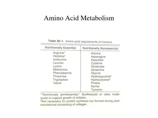
PROTEIN AND AMINO ACID METABOLISM. Prof. Dr. Mohamad Sadikin Department of Biochemistry University of Indonesia . Amino acid and Protein. Function of amino acids : 1. “Building block” of protein 2. Precursor of other biologically active compounds 3. Energy source.

E N D
PROTEIN AND AMINO ACID METABOLISM Prof. Dr. MohamadSadikin Department of Biochemistry University of Indonesia
Amino acid and Protein • Function of amino acids : 1. “Building block” of protein 2. Precursor of other biologically active compounds 3. Energy source
As Protein Building Block • There are tenth of thousand proteins in all life being • Until now: all of these enormous type of proteins consists of 2 amino acids only ( one of life being universalism) • Therefore, the differences of each protein is determined by : 1. The number of each amino acid in a given protein 2. The position of an amino acid in a protein - Both are determined by gen
Remember : • Any protein can be synthesized by a cell if the cell has a gen encoding this protein • Protein synthesis occurs only in cell (until now, protein can not be synthesized without an cell’s aid) • Most of proteins synthesized by any cell rest and work within cell
However not all of genes in genome are expressed in any given time / moment because : • Protein synthesis is very complicated and very high energy cost process • Protein is large molecule whereas intracellular space is very limited • As a highly hygroscopic large molecule, protein retains a large number of water molecule and will increase the cell size. But as the cell size is limited, protein in great concentration increases cytoplasm viscosity
Therefore : • Not all genetic information in genome will expressed in any given time, some are always expressed ( constitutive protein), some are expressed if needed ( inducible protein) and some are suppressed continually, after expressed in earlier moment of cell life ( phenomenon of cell differentiation) • Consequently, and especially for inducible protein, there are long life proteins, intermediate life proteins and short life proteins intracellular protein turnover
Intracellular protein turnover include anabolism (in this case: protein synthesis which is always an intracellular process) and catabolism ( in the protein context: intracellular protein degradation) • Anabolism in form protein synthesis is discussed in molecular biology • Intracellular protein degradation is discussed in protein and amino acid metabolism
Intracellular Protein Degradation • Eukaryotic cell has at least 3 protein degradation mechanism • Two mechanism represent by specific organelle (lysosom) or subcellular particle (proteasom) • The 3rd mechanism has no any subcellular structure and found in cytoplasm • All system contain proteolytic enzymes • All system produce free amino acids
Lysosome • A large organelle, has the same size as mitochondrion • Has all type of enzyme from the hydrolase class (protease, esterase, glycosidase, phosphatase, sulfatase) • Lysosome degrades any foreign body or substance which enters the cell through phagocytosis process usually for degrading extracellular or foreign proteins very important for cellular and body defense
All lysosomal proteases are named as cathepsin • There are several groups of cathepsin (cathepsin A, B, C and D) • As other lysosomal enzymes, all cathepsin active at acidic pH (about 5) • Cathepsin degrades protein into free amino acid
Proteasom • Subcellular particle smaller than ribosom (about 20s) • Consists of several proteases which arranged in a tube form with a lumen • Protein to be degraded is intracellular protein: 1. Misfolded proteins 2. Proteins synthesized incorrectly 3. Short life proteins
Short life protein usually is recognized by a certain amino acid sequence • Two other substrate are recognized by its unusual 3D structures • The target protein first is labeled by binding a polypeptide : ubiquitin(8,5 kD, 76 aa) • The process is catalyzed by ubiquitintransferase, a reaction which needs ATP (a synthesis reaction) • The ubiquitin part of the complex leads the ubiquitinilized protein into the lumen of proteasom and all protein degraded into aa
Calcium Dependent Papain (Calpain) Proteolytic System • Found in 1960 • Consists of calmodulin like peptide (bind Ca+ for its activity) and papain like peptide, act as cystein-protease • Recognizes the target by 3D structure • Recognizes also peptides as target through small hydrophobic aa at position 2 and large hydrophobic aa at position 1 • Needs neutral pH (unlike cathepsin) • Regulate various cellular functions by hydolize involved protein
Caspase System • Caspase (Cystein dependent aspartate directed protease) another intracellular proteolytic mechanism • The only function : activates proteins in apoptosis mechanism, by proteolysis activities
Amino Acids Metabolism • Free aa from : A.Digested food protein B.Degraded intracellular protein through various ways (lysosome, proteasom or calpain) • Will undergo: 1.Incorporation into a protein 2.Oxidation to produce ATP 3.Modification into various biologically important compound (hormone, mediators, melanin) 4. Precursor of hem, purin and pyrimidin
Oxidation of Amino Acids • Amino acids, as backbone of protein, share the same structure with 2 other major nutrients, carbohydrates and lipids • All of them have carbon skeleton (-C-C-C- ) with H and O bound directly or indirectly to C skeleton. • However, all aa have another atom bound directly to C skeleton : N, the origin of the name amino (-NH2) and in certain aa, S • Theoretically, protein can be oxidized to give rise energy
In fact, protein can be oxidized (after degraded into aa), especially in the short supply of carbohydrate and/or lipid • One condition : N (and in certain case also S) should be eliminated from the carbon skeleton • Once N and S removed, the rest carbon skeleton, because of the structural similarities with intermediates metabolites of COH and lipid, can be treated in one of the intermediate metabolism pathway of COH or lipid common or integrated metabolic pathway
It can be said that for aa carbon skeleton, there is no special or specific metabolic pathway • After N and S removing, some aa carbon skeleton resemble COH intermediate metabolite, whereas other resemble lipid intermediate metabolite • From this fact, aa can be classified into 3rd classification : - Glucogenicaa - Keto/lipogenicaa
Comprehension of the fate of any aa (glucogenic or ketolipogenic) is very important in nutrition, especially in parenteral feeding, partial or total. • Understanding of this point is also important in planning nutrition for certain diseases, as diabetes mellitus, liver diseases and kidney diseases, especially in renal failure. • As the intermediates of 3 major nutrient is interconvertible, the integrated metabolic pathway explain also how a non essential amino acid can be synthesized
Nitrogen Removing and Urea Cycle • N is removed from aa by : 1. Transamination , enzyme : aminotranferase (=transaminase) -2.Oxidative deamination, enzyme : monoamine oxidase (MAO) • In both reactions, -NH2 is removed : • In 1) : –NH2 is transferred to a -ketoacid - a new aa and a new -keto acid are formed
The reaction need also vit B6 in pyridoxal phosphate form as a transient –NH2 acceptor • The most common and most important -ketoacid as common –NH2 acceptor is -ketoglutaric acid • 2 well known with wide role aminotransferases: - Glumate-oxaloacetatetransaminase (GOT)=aspartatetransaminase (ASAT/AST) - Glutamate-pyruvatetransminase (GPT)=alanintransaminase (ALAT/ALT)
In 2) : finally, glutamic acid can reject the –NH2 and oxidized to -ketoglutaric acid. • Some aa can undergo the same reaction without transfer their –NH2 to an -keto acid. This is an oxidative deamination reaction and catalyzed by monoamine oxidase (MAO). • In some psychiatric condition, especially in depression state, there is high activity of MAO. As the level of primary amine (aa derivate) in brain is important to maintain the mental state, abnormally high active MAO is inhibited by MAO inhibitor as antidepressant.
The Fate of NH2 • The amino group removed from aa by oxidative deamination may undergo 3 ways : • Converted to –NH3 and excreted as such directly : especially in fish and tadpole - But in terrestrial animal, whose respiratory apparatus is not in direct contact with water,-NH3 is very toxic especially for CNS - For this reason, brain has a special mechanism for protecting itself against high [NH3]: glutamine synthetase, forming glutamin from glutamic acid and –NH2
Urea Cycle/Synthesis (Krebs-Henseleit Cycle) • -NH3 is very toxic, whereas glutamine synthetase is limited in the brain. • Moreover, glutamine itself is very useful (1 of 20 protein forming aa) and not a waste product • -NH3 must be rejected in the form of relatively non toxic and inert compound • For most of terrestrial animal, in the form of urea
If transamination occurs in each tissue, oxidative deamination (-NH3 release) occurs in kidney and liver, urea synthesis (=NH3 transformation and detoxification) occurs only in liver • The process begin with 1. Formation of carbamoyl phosphate: - The rx needs : carbamoyl phosphate synthetase 1 (key enzyme in urea synthesis), NH3, CO2 , ATP and O2 - Intact mitochondrion • Rx : CO2 + NH3 + 2ATP→H2N-CO-(P) + Pi
Rx 2. Citrulin synthesis. Carbamoylmoeity is transferred to aaornithin [H2N-CH2 –(CH2)2- HCNH2-COOH] & Pi is released • E : ornithintranscarbamoylase, found only inhepatocyte mitochondria • Rx 3. Argininosuccinate synthesis. NH2 of aspartate & C=O ureido of citrullin is bound each other → argininosuccinate • Need : • Argininosuccinatesynthetase and ATP • The equillibrium of rx goes to the right (argininosuccinate formation)
Rx 4. Cleavage argininosuccinate→ arginin + fumaric acid - E: argininosuccinase, found in liver and kidney • Rx 5. Releasing urea from arginine. - HN=C(NH2)NH-(CH2)2-HCNH2-COOH + H2O → (NH2)2–C=O (urea) + H2N-CH2 –(CH2)2 –H2NCH-COOH (ornithin) • E: arginase, mainly in liver. (very small qttybrain,skin,breast,kidney) • At the end of all process, urea is released and ornithin reappears and the process begin → cycling process → urea cycle / Krebs-Henseleit Cycle
Arginine Synthesis: The Urea Cycle N-Acetylglutamate synthase CoASAc Glutamate N-Acetylglutamate Activates 4 Steps Carbamoyl phosphate CPS-I NH4+ + HCO3-NH2CO2PO3-2 Ornithine Ornithine Transcarbamoylase (OTC) (mitochondria) Citrulline Ureido group
The Urea Cycle (Contd.) Asp Citrulline Arginosuccinate synthase Arginosuccinate Ornithine Transcarbamoylase (mitochondria) Fumarate Urea H2NCONH2 Argino- succinase Ornithine TCA Cycle Arginase Arginine
Fate of Carbon Skeleton • Most of carbon skeleton of aa (after removing –NH2) are also intermediate metabolite of carbohydrate (glucose) or fatty acid, especially the intermediates found post pyruvate pathway (COH) or post acetyl CoA pathway • Therefore, the aa carbon skeleton can be metabolized in glucose metabolisc pathway or fatty acid metabolic pathway or even in both pathway
These facts explain : • How non-essential aa synthesized (aided by transamination) • How protein (in the form of aa) can be used as energy source, especially in COH and lipid defficiency • That in essential, the 3 major nutrients are inter convertible each other • & consequently, all 3 major nutrient should be exist in certain balance to assure healthy metabolism process
For aromatic or cyclic side chain aa, the aromatic or cyclic structure should be opened • It is often that this process dicarboxylicketo acid as -ketoglutaric acid or oxaloacetic acid • If the ring structure contains N, the opening process produce glutamic acid or aspartic acid • 2 aa contain S (methionin and cystein). The S atom can be removed before or after –NH2 removing • S will be oxidized to –SO4 & become an essential part of glucosaminoglycan (GAG) as chondroitin sulfate and hyaluronic acid