Download
1 / 29
330 likes | 642 Views
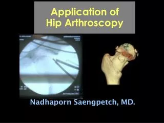
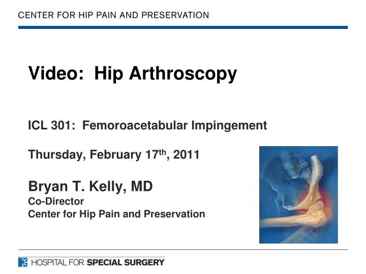
Video: Hip Arthroscopy. ICL 301: Femoroacetabular Impingement Thursday, February 17 th , 2011 Bryan T. Kelly, MD Co-Director Center for Hip Pain and Preservation. Bryan T. Kelly, MD Hospital for Special Surgery

E N D
Video: Hip Arthroscopy ICL 301: Femoroacetabular Impingement Thursday, February 17th, 2011 Bryan T. Kelly, MD Co-Director Center for Hip Pain and Preservation
Bryan T. Kelly, MD Hospital for Special Surgery Disclosure: I DO NOT have a financial interest in any commercial products or service presented in this lecture AND DO NOT INTEND to discuss off label or investigational use of products or services.
Types of financial relationships and the companies with whom I have relationships are as follows: Pivot Medical, Inc.: Consultant Smith & Nephew: Educational Consultant A2 Surgical: Consultant
Arthroscopic FAI • Set up • Access • Capsule Cut • Rim Prep / Resection • Labral Refixation • Cam Decompression • Capsular Repair
1. Patient Set Up • Adequate traction requires approximately 10mm of distraction across the joint. • Careful attention to padding is critical.
2. Access – Portals • Anterior • Anterolateral • Posterolateral Greatest Risk →→ Anterior Portal • Avg. 3 mm from a branch of the lateral femoral cutaneous nerve Primal Pictures Limited
2. Access: Expanded Portal Placement • Palpate and Outline: • Greater Trochanter • Anterior Superior Iliac Spine (ASIS) • Portal Placement • Anterolateral Portal (AL) • 1cm superior and anterior to GT • Posterolateral (PL) • 1cm superior and posterior to GT • Anterior Portal (AP) • In line with AL portal • 1 cm lateral to ASIS • Mid-Anterior Portal (MAP) • Proximal Mid-Anterior Portal (PMAP)
Portal Safety • The Mid-Anterior and Anterior portals pass in close proximity to a small terminal branch of the ascending LCFA • Greatest risk still comes from the proximity of the anterior portal to the LFCN • A slightly more lateral location may provide some protective benefit
Safe ZoneRobertson et al, Arthroscopy 2008. • The findings from this study seem to support the concept of a relative neurovascular safe zone for arthroscopic access to the hip joint within the outlined parameters.
2. Access / Visualization Transition zone injury Contra-Coup injury
3. Capsule Cut – IA Evaluation Cam Injury Rim Injury Capsular sided injury to the labrum / capsule against the rim lesion • Cam delamination • Loss of normal attachment of labrum to transition zone.
4. Rim Preparation Rim Exposure Rim Decompression Outline the rim lesion prior to decompression • Severe rim inflammation around the rim lesion
Reposition patient and fluoro for peripheral compartment work.
Pre and post fluoro shots of a patient with primary cam impingement
Pre and post fluoro shots of a patient with combined subspine / rim / and cam impingement