Download
1 / 10
100 likes | 127 Views
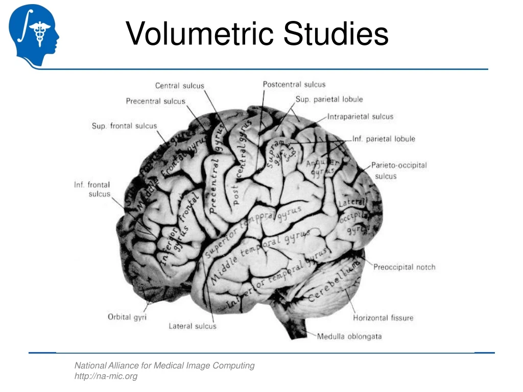
Explore the latest techniques in brain volumetric studies utilizing a 1.5 Tesla GE scanner, including image acquisition, rigid registration, brain segmentation, manual segmentation, statistical analysis, and findings on different brain regions. Discover the technical challenges and global shape measures in this field.

E N D
Current Technology • Image Acquisition: • 1.5 Tesla GE scanner, 1 coronal SPGR, 1 axial Double Echo PD and T2w image. • Rigid Registration: • Mutual Information Alignment. • Brain Segmentation: • Expectation Minimization Segmentation in 4 tissue classes (White, Gray, CSF and Face). • Manual Segmentation of Regions Of Interests. • Statistical Analysis of the ROIs’ Volume.
Brain Segmentation W. Wells
Posterior Superior Temporal Gyrus (STG)Red = Left, Green= Right• Amygdala. Yellow = Left, Light Blue = Right• Hippocampus (Hipp.). Orange = Left, Dark Blue = Right • Parahippocampal Gyrus (PHG). Dark Pink = Left, Purple = Right L. Post. STG L. STG Hipp. PHG L. Amygdala. L. Ant. STG L. Post. STG L. Hipp. Hirayasu et al.
Volumetric Studies Findings • Lateral Ventricles & CSF Enlargement. • Parietal Volume Reduction. • Temporal Lobe Volume Reductions • Amygdala-Hippocampal Complex, Parahippocampal Gyrus, & Superior Temporal Gyrus. • Prefrontal Volume Reduction. • Basal Ganglia (Caudate, Putamen, Globus Pallidus) increases and decreases.
Shape: Global Measures J. Levitt and C.F. Westin
Shape: Surface Based Analysis H.J. Park and J. Levitt
Shape: Medial Representations J. Levitt and S. Bouix
Technical Challenges • Image Quality and Acquisition Sequences. • Multi Channel Atlas Based Segmentation. • Multi Channel Image Registration. • Model Based Segmentation. • Shape Analysis. • Cortical Thickness.