Download
1 / 1

E N D
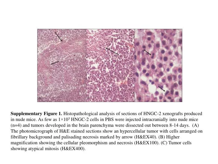
Supplementary Figure 1. Histopathological analysis of sections of HNGC-2 xenografts produced in nude mice. As few as 1×10³HNGC-2 cells in PBS were injected intracranially into nude mice (n=4) and tumors developed in the brain parenchyma were dissected out between 8-14 days. (A) The photomicrograph of H&E stained sections show an hypercellular tumor with cells arranged on fibrillary background and palisading necrosis marked by arrow (H&EX40). (B) Higher magnification showing the cellular pleomorphism and necrosis (H&EX100). (C) Tumor cells showing atypical mitosis(H&EX400).