Download
1 / 40
410 likes | 426 Views
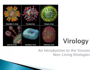
Dive into the realm of virology with this detailed guide covering virus structure, replication, detection methods, and viral life cycle. Learn about DNA and RNA viruses, viroids, prions, and lab diagnosis techniques.

E N D
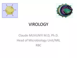
Definition of virus • Smallest infectious agent with a size ranging from 20 to 300 nm, • genome, a single nucleic acid either DNA or RNA, but never both.
Morphology of viruses • Size: 20 – 300 nm. • Filterable – ability to pass through the filters that hold back bacteria. • Ultramicroscopic – too small to be seen under the light microscope. (Except poxviruses which can be seen under the light microscope when suitably stained).
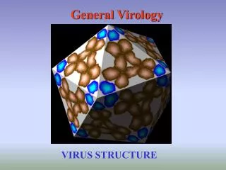
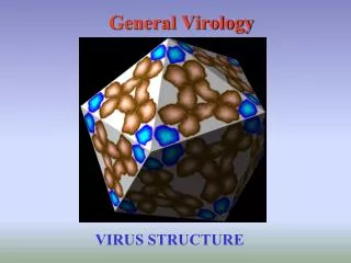
Shape - Capsid symmetry • Icosahedral (Cubic) - polygon with 12 vertices and 20 facets in the shape of equilateral triangle. E.g.; Papova, picorna and adenoviruses
Helical -Capsomeres and nucleic acid are wound together to form a helical or spiral tube. E.g.: Rabies virus, Influenza virus
Complex - Symmetry is complex and not fully understood. E.g: Pox viruses
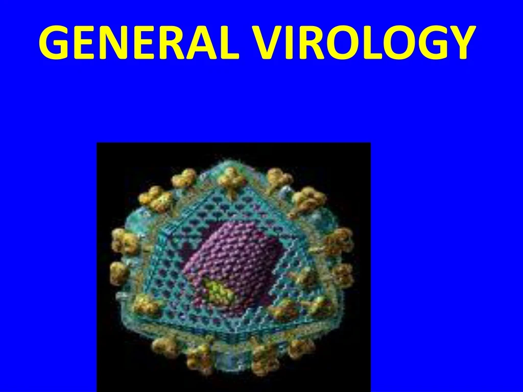
Virus structures Naked icosahedral Naked helical Enveloped icosahedral spikes (glycoprotein) capsomer protomer envelope nucleic acid (protein, lipid) Enveloped helical
Viral envelope: Viruses may be enveloped or non – enveloped. • Envelope is the outer covering of the viruses derived from the host cell membrane when the progeny virus is released by budding. • Envelope is a lipid bilayer with virus encoded proteins on the surface.
Protein subunits may be seen as projecting spikes on the surface of the envelope, which are called peplomers(peplums – envelope). • A virus may have more than one type of peplomers. • Influenza – Haemagglutinin (HA) & Neuraminidase (NA).
REPLICATION OF VIRUSES • Adsorption or Attachment • Penetration • Uncoating • Biosynthesis of viral Nucleic acid & protein • Assembly / Maturation • Release
Click after each step to view process VIRAL LIFE CYCLE ATTACHMENT HOST FUNCTIONS PENETRATION UNCOATING Transcription Translation REPLICATION ASSEMBLY (MATURATION) RELEASE MULTIPLICATION
DNA VIRUSES Parvo Papova Adeno Herpes Pox Hepadna RNA VIRUSES Picarno Calici Reo Arbo Toga Flavi Arena Corona Retro Bunya Orthomyxo Paramyxo Rhabdo
Viroids: Single stranded circular RNA molecules that lack a protein coat. They are plant pathogens • Prions:They are infectious agents without any nucleic acid. • They are highly resistant to heat, UV rays and nucleases. • They cause slow infections with long incubation period. • Example for prion diseases – Scrapie of sheep, spongiform encephalopathy, Kuru and Creutzfeldt-Jakob disease.
Electron Microscopy • viruses growing and multiplying in cell cultures can be detected using electron microscope. • Viruses may be detected in the following specimens. • Feces - Rotavirus, Adenovirus, Norwalk like viruses, Astrovirus, Calicivirus • Vesicle Fluid – HSV, VZV • Skin scrapings - papillomavirus, orf, molluscum contagiosum
Electronmicrographs Adenovirus Rotavirus
Immune Electron Microscopy • The sensitivity and specificity of EM may be enhanced by immune electron microscopy. • There are two variants:- • Classical Immune electron microscopy (IEM) • Solid phase immune electron microscopy (SPIEM)
Cultivation and detection of viral growth • Three methods are employed for cultivation of viruses. • Animal Inoculation. • Embryonated egg Inoculation. • Tissue Culture.
Embryonated Egg Inoculation • Embryonated hens eggs (7 – 12 days old) are inoculated by several routes such as – • Chorioallantoic Membrane (CAM) – used for growing mainly pox viruses and herpes viruses. • It produces visible lesions called pocks . Pocks produced by different viruses have different morphology.
Amniotic Cavity – used for primary isolation of influenza virus. • Allantoic Cavity – used for growing influenza virus for vaccine production. • Yolk Sac – used for cultivation of certain Arboviruses and bacteria likeChlamydia and Rickettsiae.
Tissue Culture • Organ Culture – small bits of organs maintained in the tissue culture growth medium can be used for isolation of certain viruses. E.g., tracheal ring culture for isolation of Coronaviruses. • Explant Culture – fragments of minced tissue can be grown as explants. This method is rarely used nowadays.
Cell Culture • Tissues are dissociated into component cells by the action of Proteolytic enzymes such as trypsin. • The cell suspension is distributed in glass or plastic tubes or Petri dishes. • On incubation, the cells divide to form a monolayer sheet of cells within a period of one week.
Viruses can be grown in living cells maintained in sterile conditions in the laboratory. • There are three types of tissue cultures.
Primary Cell cultures • These are normal cells, freshly taken from the organs of animal or human beings and cultured. • They are capable of only 5 – 10 divisions and employed for primary isolation of viruses and vaccine production. • E. g. – monkey kidney cell; human amnion cell; chick embryo fibroblast cell cultures.
Diploid cell Cultures • These are cells of a single type that contain same number of chromosomes as the parent cells and are diploid. • They can be sub-cultured up to 50 times. E. g. WI 38 (Human Embryonic Lung cell Line).
Continuous Cell Cultures • These are cells of single type that are capable of indefinite growth in vitro. • These are usually derived from cancerous tissue. • They grow faster and have haploid number of chromosomes.
These cells can be serially cultivated indefinitely and hence, are called as continuous cell lines. • These can be stored at – 70.C for use whenever necessary. • E. g. HeLa cell Lines (Human carcinoma of cervix cell lines); HEp2 cell lines (Human Epithelioma of larynx cell lines); Vero cell lines (Vervet monkey kidney cell lines).
Serology • Detection of rising titers of antibody between acute and convalescent stages of infection, or the detection of IgM in primary infection • Following serological methods can be used • Complement fixation test • Haemagglutination inhibition test • Immunofluorescence • Neutralization test • Radioimmunoassay • ELISA • Latex agglutination test • Western blot assay
Enzyme Linked Immunosorbent Assay (ELISA) Uses : • Detection and quantitation of antibodies in a wide variety of bacterial, viral and parasitic diseases like TORCH syndrome. • Detection and quantitation of antigens for the diagnosis of infectious diseases e. g. rotavirus, HBsAg. • Detection and quantitation of tumor markers e. g. AFP , CEA.
Western Blot HIV-1 Western Blot • Lane1: Positive Control • Lane 2: Negative Control • Sample A: Negative • Sample B: Indeterminate • Sample C: Positive
Molecular Methods • Methods based on the detection of viral genome are also commonly known as molecular methods. • Dot-blot, Southern blot, in-situ hybridization are examples of classical techniques. They depend on the use of specific DNA/RNA probes for hybridization. • Detection of virus genetic material (PCR, RTPCR,