Download
1 / 46
460 likes | 525 Views
Learn about the heart's structure, valves, circulation, sounds, cardiac cycle, and electrical conduction system in the cardiovascular system. Discover how the heart's chambers function in systole and diastole to pump blood efficiently through the body. Explore EKG monitoring and autonomic innervation's role in regulating heart function.

E N D
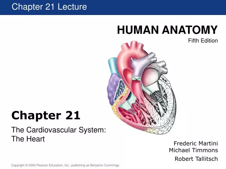
Chapter 21 Lecture Chapter 21 The Cardiovascular System: The Heart Frederic Martini Michael Timmons Robert Tallitsch
Introduction • The blood must stay in motion to maintain homeostasis. • The heart keeps blood moving. • The volume of blood pumped by the heart can vary widely, between 5 and 30 liters per minute.
An Overview of the Cardiovascular System • The heart is a small organ; it is roughly the size of a clenched fist. • The heart has four muscular chambers: • Right and left atria • Right and left ventricles
Fig 21.6
An Overview of the Cardiovascular System [Insert fig 21.1] Figure 21.1 The Pulmonary and Systemic Circuits
Orientation of the Heart Figure 21.4 Position of the Heart
The Pericardium Figure 21.2a,b The Location of the Heart in the Thoracic Cavity
The Pericardium Figure 21.2c The Location of the Heart in the Thoracic Cavity
The Pericardium Figure 21.2d The Location of the Heart in the Thoracic Cavity
The heart wall • Parietal pericardium • Epicardium/visceral pericardium • Myocardium-cardiac muscle tissue • Endocardium- • epithelia tissue Fig 21.3
Intercalated discs & gap junctions • Intercalated discs hold adjacent cardiac muscle cells together • Cells work together during contraction • Mechanically links cells together • Gap junctions allow ions to pass from cell to cell • Electrical stimulation in one cell can pass directly into other cells • Electrically/chemically links cells together • Cardiac muscle cells work as a well organized unit
Structure of the Heart Wall Figure 21.3c-e Cardiac Muscle Tissue
Fig 21.5
Fig 21.5
Fig 21.6
Internal Anatomy and Organization of the Heart Figure 21.6d Horizontal Section of Heart
Heart valves • Heart valves allow blood to flow only in one direction thru the heart • Atrioventricular valves (AV)-between an atrium and ventricle • rt. atrium>Rt. AV (tricuspid) valve>rt.ventricle • lt. atrium>Lt. AV (bicuspid/mitral) valve>lt. ventricle
The Structure and Function of Heart Valves Figure 21.7a Valves with Ventricles Relaxed
Semilunar valves-between the ventricle & an artery • lt.ventricle>Aortic semilunar valve>aorta • rt.ventricle>Pulmonary semilunar valve>pulmonary artery
The Structure and Function of Heart Valves Figure 21.7b Valves with Ventricles Contracted
Coronary Circulation Figure 21.9a Anterior Heart Figure 21.9b Posterior Heart
Pulmonary circuit - from heart to lungs back to heart Systemic circuit - from heart to body back to heart
Atria vs. ventricles • Blood enters the heart via atria • Atria have thinner walls than ventricles • Atria pump blood to the ventricles • Ventricles pump blood thru the pulmonary and the systemic circuit
Right vs. Left ventricle Fig 21.6 The left Ventricle has a much thicker myocardium
Heart sounds The two heart sounds are: • “Lub”-AV valves closing • “Dub”-semilunar valves closing • Aortic-2nd intercostals space (Right side) • Pulmonary- 2nd ICS (Left side) • Right AV valve- 5th ICS (Right of sternum) • Left AV valve- 5th ICS (inferior to left nipple)
Heart Valves and Heart Sounds • Closure of the AV valves create the 1st heart sound (‘lub’). • Closure of the semilunar valves create the 2nd heart sound (‘dub’). • Placement of a stethoscope varies depending on which heart sounds and valves are of interest.
The cardiac cycle • A chamber of the heart can be in one of two phases: • Systole-contraction of the muscle, ejecting blood out of the chamber • Diastole-relaxation of the muscle, the chamber fills with blood • The heart pumps by using cycles of systole and diastole
Cardiac Cycle • Systole: contraction phase • Diastole: relaxation phase Mid-to-late diastole . dub lub Early diastole Ventricular systole
Nodal cells • Nodal cells spontaneously depolarize causing an action potential • Two groups of nodal cells: • Sinoatrial (SA) node-makes 80-100 AP/min • Primary pacemaker • Posterior wall of the rt. atrium • Atrioventricular AV node-slower than SA node • Secondary pacemeker • Inferior region of the rt. Atrium wall
Electrical Conduction System 1. Sino Atrial (SA) Node 2. Atrial Ventricular (AV) Node 3. AV Bundle (Bundle of His) 4. L and R Bundle Branches 5. Purkinje Fibers
The Cardiac Cycle Figure 21.11 The Cardiac Cycle
Fig 21.12 The electrical signal stimulates contraction of the chambers
Figure 49.4 The Cardiac Cycle Left atrium Right atrium Right ventricle Left ventricle Ventricle contracting Ventricle relaxing P (mm Hg) V (ml) Pressure in left ventricle Left ventricular volume Pressure in aorta
EKG-electrocardiogram • Surface electrodes can monitor the depolarization of the nodal and conducting fibers • EKG graph gives electrical and mechanical diagnostic information
The Autonomic Innervation of the Heart • The stimulus for contraction is generated by pacemaker cells of the SA node. • Modified by the ANS • Modified by Hormones
Autonomic Control of Heart Rate • Basic rate established by pacemaker cells that inside the heart (myocardium) – called “intrinsic myogenic control” • Modified by ANS • Parasympathetic: ACh decreases rate and contraction force via the Vagus nerve X • Sympathetic: NE increases heart rate and force of contraction via nerve.
Cardiac Centers in CNS • Cardioaccelatory center • Medulla oblongata (Activates sympathetic neurons) • Cardioinhibitory center • Medulla oblongata (Parasympathetic neurons) Centers receive input from • Higher centers (cerebrum) • Receptors monitoring blood pressure • Receptors monitoring dissolved gases
Superior/Inferior Vena Cava • Rt. Atrium • Rt. Atrioventricular valve • Rt. Ventricle • Pulmonary Semilunar valve • Pulmonary Arteries • Lungs • Pulmonary Veins • Lt. Atrium • Lt. Atrioventricular valve • Lt. Ventricle • Aortic Semilunar valve • Ascending Aorta