Download
1 / 40
470 likes | 1.32k Views
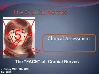
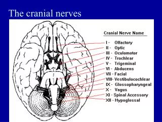
The Cranial Nerves. DR. S. H. KHAN. Names of cranial nerves. I Ⅰ Olfactory nerve II Ⅱ Optic nerve III Ⅲ Oculomotor nerve IV Ⅳ Trochlear nerve V Ⅴ Trigeminal nerve VI Ⅵ Abducent nerve VII Ⅶ Facial nerve VIII Ⅷ Vestibulocochlear nerve

E N D
The Cranial Nerves DR. S. H. KHAN
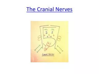
Names of cranial nerves • I Ⅰ Olfactory nerve • II Ⅱ Optic nerve • III Ⅲ Oculomotor nerve • IV Ⅳ Trochlear nerve • V Ⅴ Trigeminal nerve • VI Ⅵ Abducent nerve • VII Ⅶ Facial nerve • VIIIⅧ Vestibulocochlear nerve • IX Ⅸ Glossopharyngeal nerve • X Ⅹ Vagus nerve • XI Ⅺ Accessory nerve • XII Ⅻ Hypoglossal nerve
OH!OH!OH!TRY TOUCH AND FEEL VERY GREAT VAMPIRE AH! HELL !!!
One Officer Of The Troops Allied Forces Visited Germany, Venus And Hungary.
Classification of cranial nerves • Sensory cranial nerves: contain only afferent (sensory) fibers • I. Olfactory nerve • II. Optic nerve • VIII. Vestibulocochlear nerve • Motor cranial nerves: contain only efferent (motor) fibers • III. Oculomotor nerve • IV. Trochlear nerve • VI. Abducent nerve • XI. Accessory nerve • XII. Hypoglossal nerve • Mixed nerves: contain both sensory and motor fibers--- • V. Trigeminal nerve, • VII. Facial nerve, • IX. Glossopharyngeal nerve • X. Vagus nerve
Olfactory nerve (I) Olfactory mucosa → Cribriform foramina → Olfactory bulb
Optic nerve (II) Ganglion cell → Optic canal → Lateral geniculate body
Vestibulocochlear nerve (VIII) Vestibular ganglionVestibular nuclei Internal acoustic meatus Cochlear ganglion Cochlear nuclei
Oculomotor nerve (III) • Main action-supplies • Superior, inferior and medial recti; inferior obliquus; levator palpebrae superioris • Sphincter pupillea and ciliary muscle • Ciliary ganglion: lies between optic nerve and lateral rectus Oculomotor nerve
Abducent nerve Accessory nerve
Hypoglossal nerve Hypoglossal nerve (XII)
Oculamotor paralysis Abducent nerve injury
Branches Ophthalmic nerve • Sensory • Leave the skull through the superior orbital fissure, to enter orbital cavity • Branches • Frontal nerve • Lacrimal nerve • Nasociliary nerve
Distribution: • Cornea • Skin of forehead, scalp, eyelids, nose • Mucosa of nose & paranasal sinues
Maxillary nerve • sensory • Leave skull through foramen rotundum • Branches • Infraorbital nerve • Zygomatic nerve • Superior alveolar nerve • Pterygopalatine nerve
Distribution: • Skin of face over maxilla • Teeth of upper jaw • Mucosa of nose, maxillary sinus and palate
Mandibular nerve • Mixed • Leave the skull through the foramen ovale to enter the infratemporal fossa • Branches • Auriculotemporal nerve • Buccal nerve • Lingual nerve • Inferior alveolar nerve • Nerve of masticatory muscles
Distribution: MOTOR: • Muscles of mastication, mylohyoid, and anterior belly of digastric SENSORY: • Skin of cheek, over mandible and side of head • Teeth and gum of lower jaw • TM joint • Mucosa of mouth and anterior 2/3 of tongue
Course: leaves skull through internal acoustic meatus, facial canal and stylomastoid foramen, it then enters parotid gland where it divides into five branches which supply facial muscles
Branches within facial canal • Greater petrosal nerve • Nerve to stapedius muscle • Chorda tympani Terminal branches within parotid gland • Temporal • Zygomatic • Buccal • Marginal mandibular • Cervical
Branches outside of facial canal • Temporal • Zygomatic • Buccal • Marginal mandibular • Cervical
Function Motor • Muscles of face ( facial expression) and scalp • Stapadius, post. Bally of digastric Sensory • Taste from ant. 2/3 of tongue, floor of mouth and palate (Chorda tympani ) Secretomotor (parasympathetic) • Submandibular and sublingual salivary glands • Lacrimal gland • Glands of nose & palate
Injury to the facial nerve Bell’s palsy (low. Motor neuron lesion)
Course: leaves the skull via jugular foramen Branches • Lingual branches : to taste buds and mucosa of posterior third of tongue • Pharyngeal branches : take part in forming the pharyngeal plexus • Tympanic nerve : fibers via tympanic and lesser petrosal nerves to otic ganglion, with postganglionic fibers via auriculotemporal to parotid gland • Carotid sinus branch : innervations to both carotid sinus and glomus • Others: tonsillar and stylophayngeal branches Otic ganglion: situated just below foramen ovale
Branches in neck • Pharyngeal • Superior laryngeal nerve: • Internal branch • External branch • Branches to carotid body • Right recurrent laryngeal n. • Cervical cardiac branches : descending to terminate in cardiac plexus
Superior laryngeal nerve Internal branch External branch
Branches in thorax • Cardiac • Left recurrent laryngeal n. • Pulmonary • esophageal
Branches in abdomen • Anterior and posterior gastric branches • Hepatic branches: join hepatic plexus and then supply liver and gallbladder • Celiac branches • Renal
khansarwar16@yahoo.com Always keep in touch