Download
1 / 106
1.25k likes | 2.06k Views
Eye Fun Facts. Most people blink every 2-10 seconds.Each time you blink, you shut your eyes for 0.3 seconds, which means your eyes are closed at least 30 minutes a day just from blinking.

E N D
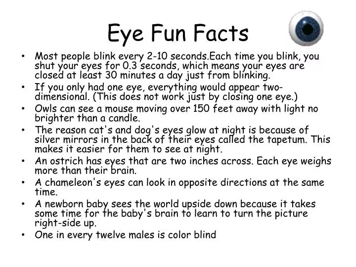
Eye Fun Facts • Most people blink every 2-10 seconds.Each time you blink, you shut your eyes for 0.3 seconds, which means your eyes are closed at least 30 minutes a day just from blinking. • If you only had one eye, everything would appear two-dimensional. (This does not work just by closing one eye.) • Owls can see a mouse moving over 150 feet away with light no brighter than a candle. • The reason cat's and dog's eyes glow at night is because of silver mirrors in the back of their eyes called the tapetum. This makes it easier for them to see at night. • An ostrich has eyes that are two inches across. Each eye weighs more than their brain. • A chameleon's eyes can look in opposite directions at the same time. • A newborn baby sees the world upside down because it takes some time for the baby's brain to learn to turn the picture right-side up. • One in every twelve males is color blind
Vision • The best-studied sense. • One quarter (1/4) of the human brain is dedicated to vision • Much of what we know is from experiments on monkeys and cats https://secure.wikimedia.org/wikipedia/commons/wiki/File:Ventral-dorsal_streams.svg
The Eye and Its Connections to the Brain • Cornea: transparent front part of the eye that covers the iris & pupil. Focuseslight. • Pupil: opening in the center of the eye that allows light to pass through • Lens: focuses the light on the retina • Retina: back surface of the eye that contains the photoreceptors • Fovea: point of centralfocus on the retina • The Route Within the Retina • photoreceptors-rods and cones • bipolar cells-receive input from rods and cones • ganglion cells-receive input from bipolar cells • opticnerve-made up of axons of ganglion cells • blindspot-the point where the optic nerve leaves the eye (called the optic disk)
Figure 17-5c The Sectional Anatomy of the Eye Visual axis Anterior cavity Cornea Edge ofpupil Anteriorchamber Posteriorchamber Iris Suspensory ligament of lens Nose Corneal limbus Conjunctiva Lacrimal punctum Lacrimal caruncle Lower eyelid Medial canthus Lateralcanthus Ciliaryprocesses Lens Ciliary body Ora serrata Sclera Choroid Retina Posteriorcavity Ethmoidallabyrinth Lateral rectusmuscle Medial rectusmuscle Optic disc Fovea Optic nerve Orbital fat Central arteryand vein Horizontal dissection of right eye
The Eye and Its Connections to the Brain • The Route Within the Retina • photoreceptors-rods and cones • bipolar cells-receive input from rods and cones • ganglion cells-receive input from bipolar cells • opticnerve-made up of axons of ganglion cells • blindspot-the point where the optic nerve leaves the eye (called the optic disk)
Figure 17-7a The Organization of the Retina Rod Horizontal cell Cone Pigmentedpart of retina Rods andcones Amacrine cell Bipolar cells Ganglion cells LIGHT The cellular organization of the retina. The photoreceptors are closest to the choroid, rather than near the posterior cavity (vitreous chamber).
Figure 17-7a The Organization of the Retina Choroid Pigmentedpart of retina Rods andcones Bipolar cells Ganglion cells LM 350 Retina Nuclei ofganglion cells Nuclei ofbipolar cells Nuclei of rodsand cones The cellular organization of the retina. The photoreceptors are closest to the choroid, rather than near the posterior cavity (vitreous chamber).
Figure 17-7b The Organization of the Retina Pigmentedpart of retina Neural part of retina Central retinal vein Optic disc Central retinal artery Sclera Choroid Optic nerve The optic disc in diagrammatic sagittal section.
Figure 17-7c The Organization of the Retina Optic disc(blind spot) Fovea Macula Central retinal artery and veinemerging from center of optic disc A photograph of the retina as seen through the pupil.
The Eye Lens Cornea Retina Pupil Light first passes through the cornea, which performs about ¾ of the focusing. wikimedia.org/wikipedia/commons/wiki/File:Schematic_diagram_of_the_human_eye.svg
The Eye Lens Cornea Retina Pupil Light must then pass through the pupil. The lens then adjusts the light and creates a clear image for the photoreceptors in the retina.
The amount of light allowed into the eye is regulated by the Pupil, controlled by the Iris. • Fine adjusting of the light is done by manipulating the shape of the lens, controlled by muscles behind the iris.
Quick Review Cornea • Transparent front part of the eye that covers the iris & pupil. Focuseslight. • Opening in the center of the eye that allows light to pass through • Focuses the light on the retina • Back surface of the eye that contains the photoreceptors • Point of centralfocus on the retina Pupil Lens Retina Fovea
The Macula • The area around the fovea, called the macula, is critical for reading and driving. • Macular Degeneration is the leading cause of blindness in the elderly population
More Review • Generic term for neural sensory receptor specific to detecting light: • The answer to number 1 is made up of two major groups of cells. The group that is associated with detecting colors are called: • The answer to number 1 is made up of two major groups of cells. The group that is associated with detecting light under dim conditions are called: photoreceptors cones rods
Even more Review. • What are the names of the cells that receive input from the rods and cones? • The answer to ‘1’ then synapses with retinal ___________ cells. • What is the optic nerve • Explain the optic disk. Bipolar cells ganglion The optic nerve relays sensory information from the retina to the brain. It is made up of the axons of the retinal ganglion cells. This is where the optic nerve exits the eye. It is also referred to as the blind spot.
Last Review, for now • Explain the route (structures involved) light takes to enter the eye and leave the eye as an electrical signal. Lens Cornea Pupil Retina Retinal ganglion cells Bipolar cells Photoreceptors Optic Nerve Photoreceptors Bipolar cells Retinal ganglion cells Out the optic disk
The Eye-Lense • The Lens • Lens fibers • Cells in interior of lens • No nuclei or organelles • Why is this important? • Filled with crystallins, which provide clarity and focusing power to lens • Cataract • Condition in which lens has lost its transparency
Light Refraction • Bending of light by cornea and lens • Focal point • Specific point of intersection on retina • Focal distance • Distance between center of lens and focal point Focal distance Focal distance Focal distance Closesource Focalpoint Light from Lens distant source (object) The rounder the lens,the shorter the focal distance The closer the light source,the longer the focal distance
Light Refraction of Lens • Accommodation • Shape of lens changes to focus image on retina • Astigmatism • Condition where light passing through cornea and lens is not refracted properly • Visual image is distorted
Quick demo 1.) Hold your pen out one foot from your face. Focus on this object and observe how your eyes feel 2.) Focus on Something Mr. O’Neil tell’s you to. (far away) Observe how your eyes feels. Under which condition (1 or 2) did your eyes feel more relaxed?
Figure 17-11 Accommodation For Close Vision: Ciliary Muscle Contracted, Lens Rounded Lens rounded Focal pointon fovea Ciliary musclecontracted For Distant Vision: Ciliary Muscle Relaxed, Lens Flattened Lens flattened Ciliary musclerelaxed
Light Refraction of Lens • Image reversal • Visual acuity • Clarity of vision • “Normal” rating is 20/20 Light from a point at the top of anobject is focused on the lowerretinal surface. Light from a point at the bottom ofan object is focused on the upperretinal surface.
Figure 17-12c Image Formation Light rays projected from a verticalobject show why the image arrivesupside down. (Note that the image isalso reversed.)
Figure 17-12d Image Formation Light rays projected from a horizontalobject show why the image arriveswith a left and right reversal. Theimage also arrives upside down. (Asnoted in the text, these representa-tions are not drawn to scale.)
Figure 17-13 Accommodation Problems A camera lens has a fixed size and shapeand focuses by varyingthe distance to the film. The eye has a fixedfocal length andfocuses by varyingthe shape of the lens.
Figure 17-13 Accommodation Problems Emmetropia(normal vision)
Myopia • Eyeball is too deep or the resting curvature of the lens is too great • Image of a distant object is projected in front of the retina. • The person will see distant objects as blurry and out of focus. • Vision at close range will be normal because the lens is able to round as needed to focus the image on the retina. Myopia Myopiacorrected witha diverging,concave lens What is the common name for Myopia? nearsightedness Diverginglens
Figure 17-13 Accommodation Problems If the eyeball is too shallow or the lens is too flat, hyperopia results. The ciliary muscle must contract to focus even a distant object of the retina. And at close range the lens cannot provide enough refraction to focus an image on the retina. Older people become farsighted as their lenses lose elasticity, a form of hyperopia called presbyopia (presbys, old man). Hyperopia (farsightedness) Hyperopiacorrected witha converging,convexlens Converginglens
Figure 17-13 Accommodation Problems Surgical Correction Variable successat correcting myopia and hyperopia hasbeen achieved by surgery that reshapes the cornea. In Photorefractivekeratectomy (PRK) a computer-guidedlaser shapes the cornea to exactspecifications. The entire procedure can be done in less than a minute. A variation on PRK is called LASIK (Laser-Assisted in-Situ Keratomileusis). In this procedure the interior layers of the cornea are reshaped and then re-covered by the flap of original outer cornealepithelium. Roughly 70 percent of LASIK patients achieve normal vision, and LASIK has become the most common form of refractive surgery. Even after surgery, many patients still need reading glasses, and both immediate and long-term visual problems can occur.
Eye Movement • There are the 6 small muscles that move each eye from side to side, up and down and on the slant. • When these muscles don't work together, it can affect vision. • “Lazy eye” is a condition that affects about 5% of children and arises when the eye muscles don't work together properly. • One eye takes over all the vision duties.
Binocular Vision • Because the two eyes are located in different positions, each takes in a unique view from its own perspective. • Visual signals from both eyes mix and match on their way to the brain • The image from each eye is not exactly the same! • Allows for depth perception
Binocular Vision • When you look at a scene with both eyes, the objects to your left register on the right side of the retina. • This visual information then maps to the right side of the cortex. • The result is that the left half of the scene you are watching registers in the cerebrum’s right hemisphere.
Pathway of Visual Signals: From the eyes to visual cortex Left Visual Field Right Visual Field In the retina, light signals are converted to electric signals. Optic nerves carry the electric signals from each eye. Optic nerves meet at the optic chiasm. Some fibers cross to the other side of the brain, while others stay on the same side. LGN LGN Lateral geniculate nucleus Right Visual Cortex Left Visual Cortex Visual cortex
STEREOPSIS • The two separate images are sent on to the brain for processing. • When the two images arrive simultaneously in the back of the brain they are united into one picture. • The combined picture appears three-dimensional (3-D) because it has the added depth dimension. • That's stereo vision. Stereo vision gives you depth perception.
Remember the 3 Layers of the Retina • The retina contains three stages of neurons: • The first (closest to the brain), the layer of rods and _____, sends its signals to: • The middle layer, which contains _______ cells & relays signals to: • The thirdlayer, which consists of the retinal __________cells whose axons form the ______ _____. cones bipolar ganglion optic nerve
Figure 6.5 Details of Retinal Circuitry Adapted from Dowling, J. E., and Boycott, B. B. Proceedings of the Royal Society of London, B, 1966, 166, 80–111.
Photoreceptors Photoreceptors absorb light and send electrical signals to nearby neurons lining the back of the eye.
Photoreceptors • 125 Million specialized neurons per eye that turn light into electrical signals. • There are two major groups: • Cone Cells • Three types (red, green, and blue) • Colorvision • Fine detail • 2. Rod Cells • Used in dim light • Sensitive to motion • Black and white sensitivity http://www.kurzweilai.net/images/Photoreceptors.jpg
Figure 17-14a Structure of Rods, Cones, and Rhodopsin Molecule Pigment Epithelium In a cone, the discs are infoldings ofthe plasma membrane, and the outersegment tapers to a blunt point. In a rod, each disc is an independententity, and the outer segment formsan elongated cylinder. Melanin granules Outer Segment Discs Connectingstalks Inner Segment Mitochondria Golgiapparatus Nuclei Cone Rods Each photoreceptorsynapses with a bipolar cell. Bipolar cell LIGHT Structure of rods and cones.
RODS & CONES • Rods • 95% of photoreceptors • abundant in the periphery of the retina • best for low light conditions • See black/white and shades of gray • Cones • abundantaroundfovea (only red & green) • best for brightlight conditions • see color • Accounts for most of human vision
Visual Physiology • Color Vision • Integration of information from red, green, and blue cones • Color blindness • Inability to detect certain colors
Figure 17-15 Cone Types and Sensitivity to Color Rods Redcones Bluecones Greencones Light absorption(percent of maximum) W A V E L E N G T H (nm) Violet Blue Green Yellow Orange Red