Download
1 / 21
210 likes | 344 Views
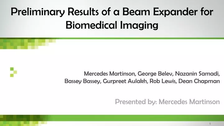
Mercedes Martinson, George Belev , Nazanin Samadi , Bassey Bassey , Gurpreet Aulakh , Rob Lewis, Dean Chapman. Preliminary Results of a Beam Expander for Biomedical Imaging. Presented by: Mercedes Martinson . Outline. Background Motivation Implementation Results Applications

E N D
Mercedes Martinson, George Belev, NazaninSamadi, BasseyBassey, GurpreetAulakh, Rob Lewis, Dean Chapman Preliminary Results of a Beam Expander for Biomedical Imaging Presented by: Mercedes Martinson
Outline • Background • Motivation • Implementation • Results • Applications • Future work
The Canadian Light Source (CLS) • Electron beam inside a storagering • Bent using dipole bend magnets • Acceleration of charged particles creates electromagnetic radiation • Dopper“headlight” effect shifts frequency higher • Pushes radiation into X-ray regime • Small vertical divergence • Horizontal “smearing”
Synchrotron beam is wide and short • Vertical scanning • CT imaging: slow • Imaging: slices • Reconstruction: stitching • Increases dose • Dynamic imaging: • Basically impossible
Double bent Laue crystal expander Expansion:
Geometric Focus (monochromatic) Curvature of crystal and lattice planes causes a virtual focus.
Single ray Focus (polychromatic) Reflection from multiple layers and angle between lattice planes causes a virtual focus of the beam
Single ray + geometric focus matched The beam appears to come from a point (virtual focus) and the beam diverges on the downstream side.
Rigid Frame Bender Advantages: • Bending radius is reproducible • Uniform bend • Anticlastic bending is reduced • Inexpensive • Heat sink Disadvantages: • Bending radius is fixed • Crystal distortion • Window height limits expansion • Machining irregularities
How much bigger did we make it? • 7.7x(Summer 2013) • Beam quality was poor: • Phase • Uniformity • Vertical blurring • 12x (Winter 2014) • Improved beam quality: phase and edge preservation • Flux reduction: low intensity reflection
What did we do with the bigger beam? • Measured beam quality • Preliminary imaging tests • High resolution micro-CT in a single spin • Full FOV on 8.75 μm Hamamatsu detector • Dynamic imaging • Live mouse, 30 fps (200 μm) • Measured flux • Comparable to beamline mono • Phase imaging
Preliminary imaging tests Various phase, absorption, and edge test objects. This was before we figured out the trick to getting a uniform beam.
In-line phase image using expander at CLS 50 mm Approximate height using beamline mono CLS BMIT-BM: Energy = 33 keV, Propagation distance = 2.05 m
Mercedes Martinson, NazaninSamadi, and BasseyBassey are Fellows , and Dean Chapman and Rob Lewis are Mentors, in the Canadian Institutes of Health Research Training grant in Health Research Using Synchrotron Techniques (CIHR-THRUST)
References • Astolfo, A., Schültke, E., Menk, R. H., Kirch, R. D., Juurlink, B. H. J., Hall, C., Harsan, L.-A., Stebel, M., Barbetta, D., Tromba, G. & Arfelli, F. (2013). Nanomedicine : nanotechnology, biology, and medicine 9, 284-292. • Bravin, A., Coan, P. & Suortti, P. (2013). Physics in Medicine and Biology 58, R1. • Coan, P., Bravin, A. & Tromba, G. (2013). Journal of Physics D: Applied Physics 46, 494007. • Erola, E., Eteläniemi, V., Suortti, P., Pattison, P. & Thomlinson, W. (1990). Journal of Applied Crystallography 23, 35-42. • Hooper, S. B., Kitchen, M. J., Siew, M. L. L., Lewis, R. A., Fouras, A., B te Pas, A., Siu, K. K. W., Yagi, N., Uesugi, K. & Wallace, M. J. (2009). Clinical and Experimental Pharmacology and Physiology 36, 117-125. • Hubbell, J. H. a. S., S.M. (2004). Tables of X-Ray Mass Attenuation Coefficients and Mass Energy-Absorption Coefficients, http://physics.nist.gov/xaamdi. • Hyodo, K., Ando, M., Oku, Y., Yamamoto, S., Takeda, T., Itai, Y., Ohtsuka, S., Sugishita, Y. & Tada, J. (1998). Journal of Synchrotron Radiation 5, 1123-1126. • Lewis, R. A. (2004). Physics in Medicine and Biology 49, 3573-3583. • Lewis, R. A., Yagi, N., Kitchen, M. J., Morgan, M. J., Paganin, D., Siu, K. K. W., Pavlov, K., Williams, I., Uesugi, K., Wallace, M. J., Hall, C. J., Whitley, J. & Hooper, S. B. (2005). Physics in Medicine and Biology 50, 5031-5040. • Liu, Y., Nelson, J., Holzner, C., Andrews, J. C. & Pianetta, P. (2013). Journal of Physics D: Applied Physics 46, 494001. • Porra, L., Suhonen, H., Suortti, P., Sovijärvi, A. R. A. & Bayat, S. (2011). Critical Care Medicine 39, 1731-1738 1710.1097/CCM.1730b1013e318218a318375. • Schültke, E., Kelly, M. E., Nemoz, C., Fiedler, S., Ogieglo, L., Crawford, P., Paterson, J., Beavis, C., Esteve, F., Brochard, T., Renier, M., Requardt, H., Dallery, D., Le Duc, G. & Meguro, K. (2011). European journal of radiology 79, 323-327. • Suortti, P., Keyriläinen, J. & Thomlinson, W. (2013). Journal of Physics D: Applied Physics 46, 494002. • Suortti, P. & Schulze, C. (1995). Journal of Synchrotron Radiation 2, 6-12. • Suortti, P. & Thomlinson, W. (2003). Physics in Medicine and Biology 48, R1. • Thomlinson, W., Suortti, P. & Chapman, D. (2005). Nuclear Instruments & Methods in Physics Research Section a-Accelerators Spectrometers Detectors and Associated Equipment 543, 288-296.