Download
1 / 80
2.12k likes | 6.14k Views
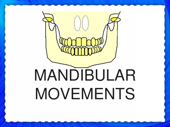
MANDIBULAR MOVEMENTS. CONTENTS. Introduction Anatomy of TMJ Muscles of Mastication Mandibular Movements Eccentric Mandibular Movements Clinical Significance of Mandibular Movements Methods Used For Recording Mandibular Movements Conclusion. INTRODUCTION.

E N D
CONTENTS • Introduction • Anatomy of TMJ • Muscles of Mastication • Mandibular Movements • Eccentric Mandibular Movements • Clinical Significance of Mandibular Movements • Methods Used For Recording Mandibular Movements • Conclusion
INTRODUCTION • The masticatory system is extremely complex. • It is made up of three linked chains.
PASSIVE MUSCLES & LIGAMENTS MAXILLA & MANDILBLE TMJ PASSIVE
During performance of various functions there is a delicate balance between various components. • It is important to study mandibular movements as it enables us to plan arrangement of teeth & selection of articulators so that artificial prosthesis is in harmony with stomatognathic system.
ANATOMY OF TMJ • TMJ is one of the most complex joints in the body. • It is called as GINGLYMOARTRODIAL JOINT. • COMPOUND JOINT. • TMJ consists of 4 main structures:- • Condyle • Temporal bone (Squamous part) • Articular disc • Ligaments
CONDYLE • It is the portion of the mandible that articulates with the cranium, around which movement occurs.
TEMPORAL BONE • The mandibular condyles articulates at the base of the cranium with the squamous portion of the temporal bone. • ARTICULAR OR GLENOID FOSSA • SQUAMOTYMPANIC FISSURE • ARTICULAR EMINENCE.
ARTICULAR DISC • Functionally – articular disc serves as a non ossified bone that permits the complex movements of the joint. SAGITTAL PLANE ANTERIOR VIEW
MUSCLES OF MASTICATION • The skeletal muscles provide for the locomotion necessary for the individual to survive. • 4 muscles make up a group called Muscles of Mastication • Masseter • Temporalis • Medial Pterygoid • Lateral Pterygoid • Digastric also plays an important role in mandibular function.
CLASSIFICATION:- I) According to Sharry:- a) According to direction - Opening and closing movements Protrusion and retraction Lateral gliding movements b) According to tooth contact - Movements with tooth contact Movements without tooth contact c) Limitation by joint structure - Border movements Intra border movements d) Functions of masticatory system - Mastication Deglutition Speech Respiration e) CNS - Innate movements – breathing & swallowing Learned movements – speech and chewing
II) According to the type of movement occurs in TMJ:- a) Rotational b) Translation III) According to the planes of border movements:- a) Sagittal plane border movement b) Horizontal plane border movements c) Frontal plane border movements
MANDIBULAR MOVEMENTS TYPES:- • 2 types of movement occur in TMJ:- • Rotational • Translational ROTATIONAL MOVEMENT:- • Rotation – “movement of a body about its axis.” • In masticatory system, rotation occurs when the mouth opens and closes around a fixed point or axis within the condyles.
HORIZONTAL AXIS OF ROTATION:- • Mandibular movement around the horizontal axis is an opening and closing motion, referred to as Hinge Movement and horizontal axis around which it occurs is therefore referred to as “HINGE AXIS”. • The Hinge movement is the only example of mandibular activity in which a “pure” rotational movement occurs. • TERMINAL HINGE AXIS When the condyles are in their most superior position in the articular fossae and the mouth is purely rotated open, the axis around which movement occurs is called the ‘Terminal Hinge Axis’.
FRONTAL (VERTICAL) AXIS OF ROTATION:- • Mandibular movement around the frontal axis occurs when one condyle moves anteriorly out of terminal hinge position with the vertical axis of opposite condyle remaining in the terminal hinge position.
SAGITTAL AXIS OF ROTATION:- • Mandibular movement around the sagittal axis occurs when one condyle moves inferiorly while other remains in the terminal hinge position.
TRANSLATIONAL MOVEMENT:- • Translation can be defined as a movement in which every point of the moving object has simultaneously the same velocity and direction. • In masticatory system it occurs when the mandible moves forward during protrusion. The teeth, condyles and rami all move in the same direction and to the same degree. • Itoccurs within the superior cavity of the joint, between the superior surface of the articular disc and the inferior surface of the articular fossa.
SINGLE-PLANE BORDER MOVEMENTS:- • When the mandible moves through the outer range of motion, reproducible and describable limits result, which are called BORDER MOVEMENTS.
SAGITTAL PLANE BORDER & FUNCTIONAL MOVEMENTS:- • They have 4 distinct movement components:- 1) Posterior opening border: determined by ligaments & the morphology of TMJ’s. 2) Anterior opening border determined by occlusal Superior contact border & incisal surfaces of teeth. 3) Functional determined by conditional responses of neuromuscular system.
Posselt’s Figure MP ICP RCP HA MP = Maximal protrusion ICP = Intercuspal position RCP= Retruded Contact position HA = Hinge axis MO = Maximum opening MO
Posterior Opening Border Movements:- • Occurs as two stage hinging movements. • 1st stage:- • Condyles are stabilized in their most superior positions in the articular fossae.( i.e.terminal hinge position). • The mandible can be lowered (i.e. mouth opening) in a pure rotational movement without translation of condyles.
In CR, the mandible can be rotated around the horizontal axis to a distance of 20-25 mm as measured between the incisal edges of maxillary and mandibular incisors. • At this point of opening, the T.M. ligaments tighten, after which continued opening results in an anterior & inferior translation of condyles.
2nd Stage:- • As the condyle translates the axis of rotation of the mandible shifts into the bodies of rami likely to be the area of attachment of sphenomandibular ligament, resulting in the second stage of the posterior opening border movement.
During this stage, the condyles move anteriorly and inferiorly and the mandible moves posteriorly and inferiorly. • Maximum opening is reached when capsular ligaments prevent further movement of the condyles. • Maximum opening range is 40-60 mm.
Anterior Opening Border Movements:- • With the mandible maximally opened, closure accompanied by contraction of inferior lateral pterygoids (which keep the condyles positioned anteriorly) will generate the anterior border movement.
Because the maximum protrusive position is determined in part by stylomandibular ligaments, when closure occurs, tightening of ligaments produces a posterior movement of the condyles. • The posterior movement of the condyle from the maximally open position to maximally protruded position produces eccentricity in the anterior border movement. Therefore, it is not a pure hinge movement.
Superior Contact Border Movements:- • Throughout this entire border movement tooth contact is present. • It depends on:- • Amount of variation between centric relation and maximum intercuspation. • The steepness of the cuspal inclines of the posterior teeth. • Amount of vertical and horizontal overlap of anterior teeth • Lingual morphology of maxillary anterior teeth. • General interarch relationships of the teeth.
The initial tooth contact in terminal hinge axis or centric relation occurs between the mesial inclines of maxillary tooth & distal inclines of mandibular tooth. • When muscular force is applied to the mandible, a superoanterior movement or shift will occur until the intercuspal position is reached.
The slide from CR to maximum intercuspation, may have a lateral component. Average distance is 1.25 ± 1 mm. • In the intercuspal position, the opposing anterior teeth usually contact. • When the mandible is protruded, from maximum intercuspation, contact between the incisal edges of the mandibular anterior teeth & lingual inclines of maxillary anterior teeth result in an anteroinferior movement of the mandible.
This continues until the maxillary and mandibular anterior teeth are in edge to edge relationship, at which a horizontal movement continues until incisal edges of mandibular teeth pass beyond the edges of maxillary teeth.
At this point mandible moves in a superior direction until the posterior teeth contact. • The occlusal surfaces of posterior teeth then dictate the remaining pathway to the maximum protrusive movement, which joins with the most superior portion of the anterior opening border movement.
Functional Movements:- • Functional movement occurs during functional activity of the mandible. They usually take place within the border movements & therefore, considered as free movements. • Most functional movements require maximum intercuspation & therefore typically begin at & below the intercuspal position.
When mandible is at rest, it is found to be located approximately 2 to 4 mm below the intercuspal position. This is called the Clinical Rest Position. • Postural position – Since, clinical rest position is not a true resting position, the position in which mandible is maintained is termed as ‘postural position.’
Chewing Stroke:- If it is examined in sagittal plane, the movement will be seen to begin at the intercuspal position & drop downward & slightly forward to position of desired opening. It then returns in a straighter pathway, slightly posterior to the opening movement.
HORIZONTAL PLANE BORDER & FUNCTIONAL MOVEMENTS:- • When mandibular movements are viewed in the horizontal plane, a rhomboid-shaped pattern can be seen that has a functional component, & 4 distinct movement components:- 1) Left lateral border 2) Continued left lateral border with protrusion 3) Right lateral border 4) Continued right lateral border with protrusion
Left Lateral Border Movements:- • With the condyles in the centric relation position, contraction of the right inferior lateral pterygoid move the right condyle - anteriorly and medially. • If left inferior pterygoid stays relaxed, with the left condyle still in the CR & result will be left lateral border movement. • Left condyle- working or rotatory Right condyle- non-working or orbiting
Continued Left Lateral Border Movements With Protrusion:- • With the mandible in the left lateral border position, contraction of the left inferior lateral pterygoid along with continued contraction of right inferior lateral pterygoid will cause the left condyle to move anteriorly to the right.
Right Lateral Border Movements:- • Left condyle- orbiting Right condyle- rotatory
Functional Movements:- • As in the sagittal plane, functional movement in the horizontal plane most often occur near the intercuspal position. • During chewing the range of jaw movements begins some distance from maximum intercuspal position; but as the food is broken down into smaller particles, jaw action moves closer and closer to intercuspal position.

![TEMPROMANDIBULAR JOINT AND MOVEMENTS MANDIBULAR [ T M J ]](https://cdn3.slideserve.com/6414015/slide1-dt.jpg)