Download
1 / 58
580 likes | 798 Views
Development. Rachel Miller. Gamete Formation. Eggs develop in female’s ovaries - female is born germ cells in ovaries begin meiosis - division stops at prophase I - ovarian cells become primary oocytes - finish meiosis with fertilization by sperm. Gamete Formation.

E N D
Development Rachel Miller
Gamete Formation • Eggs develop in female’s ovaries - female is born germ cells in ovaries begin meiosis - division stops at prophase I - ovarian cells become primary oocytes - finish meiosis with fertilization by sperm
Gamete Formation • Sperm develop in male testes - begins at puberty - spermatogenesis takes 64 days - several million sperm produced per day
Incorrect Gametogenesis can Cause: • Down’s Syndrome • Klinefelter’s Syndrome • Edwards Syndrome • Spontaneous abortion
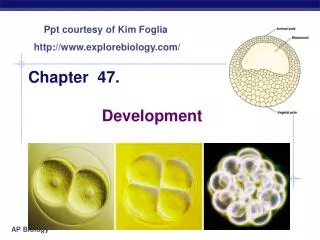
Embryonic Development • Fertilization • Cleavage • Morula • Blastula • Gastrula • Extraembryonic Membrane Development
Fertilization • Recognition - sperm secretes protein - binds with special receptor molecule on the zonapellucida - zonapellucida- glycoprotein membrane surrounding oocyte - insures fertilization between same species
Fertilization 2) Penetration - sperm and oocyte plasma membranes fuse
Fertilization 3) Fertilization membrane forms to block any more sperm
Fertilization 4) Oocyte undergoes meiosis II - produces an ovum and polar body - polar body released
Fertilization 5) Sperm and ovum nuclei fuse - zygote nucleus of 23 chromosomes
Cleavage • Rapid cell divisions without cell growth • Microfilaments contract and pinch cell in two • Form smaller blastomeres
Cleavage • Cytoplasmic Localization • Blastomeres receive different maternal messages • Inherit different proteins • Determines later cell lineages
Mammal Cleavage • Embryos genome produces proteins for cleavage to occur • Mammals take 12-24 hours for each cut • Mouse and goat- maternal to zygotic control at two cell stage • Have rotational cleavage
Day 4: Morula • Ball of 16- 32 cells
Blastula • Liquid fills morula and pushes cells out • Hollow, single layer of cells
Day 5: Blastocyst • Inner cell mass- few cells that will become actual embryo • Zonapellucida • Trophoblast- develop into placenta • Blastocoel- fluid filled cavity
Days 6-7: Blastula Hatches • Blastocyst adheres to uterine lining • Injected out of ZonaPellucida • Has now formed connections with mother’s tissues
Ectopic Pregnancy • Fertilized egg implants outside of uterus • Typically occurs in fallopian tubes • Fetus will burst organ
Days 10-14: - Fluid filled amniotic cavity opens up - Amnion forms- will enclose embryo - buoyant cradle and nourishment -Yolk sac starts to form- will make blood and germ cells - Embryo starts to form from embryonic disc - Chorion (placenta) starts to form
Days 10-14 1. Start of the embryonic disc 2. Amniotic cavity- filled with fluid -buoyant cradle and nourishment 3. Yolk sac- gives rise to blood and germ cells 4. Chorion (placenta) starts to form 4
Day 15: Gastrula • Single layered blastula becomes double layered • Opening to outside
Day 15: Gastrula • 3 Germ Layers for tissue to develop from - Ectoderm, Mesoderm, Endoderm • Archenteron- center cavity • Blastopore- opening
Days 18-23: Morphogenesis • Organs begin to form • Tissues fold • Neural folds merge to make start of spinal cord and brain • Somites- will become bones and dermis
Day 24-25 Pharyngeal arches will become: - face - neck - mouth - nasal cavities - larynx - pharynx
Differentiation • All cells started out with same genes (from one fertilized egg) • All transcribe many of same genes • Most have special functions - Ex. red blood cells transcribe genes for hemoglobin • Over time specific subsets of genes have been turned on
Inductive Cell from One Group Influences Development of Another
Pattern Formation • Sculpting of specialized tissues and organs from clumps of cells in proper places in embryo in proper order • Embryonic cell reads it genes • Makes and secretes molecules to neighbors • These signals induce changes
Cadherin and Integrin Signals • Proteins that affect how cells link up in gastrula - will affect how organs form • Signals activate/ inhibit adhesion proteins
HOX Genes • Class of master genes that contain information about mapping out of body plan
HOX Genes • All body appendages start out as tissue buds • Bud forms where Dll gene is expressed - turned on- arm/leg forms - turned off- nothing • Hox genes suppress DII when needed • DII expressed similarly across phyla
Fate Mapping • shows how a cell or tissue moves and what it develops into
Polymelia • Results in extra limb(s) - usually shrunken/deformed
Conjoined Twins • normal identical twins- the egg splits at four to eight days after fertilization • conjoined twins- the split occurs sometime after day 13 • Embryos do not fully separate
Rare Types • Dicephalus: Twins that share one body, but have two separate heads and necks. • Parasitic twins: This occurs when one smaller, malformed twin is dependent on the larger, stronger twin for survival. • Fetus in fetu: In this unusual case, one fetus grows inside the body of the other twin "Conjoined Twins Information on Healthline." Medical Information for Healthy Living | Healthline. Web. 14 Apr. 2011. <http://www.healthline.com/galecontent/conjoined-twins/2>.
http://www.metacafe.com/watch/122740/baby_born_with_2_heads/