Download

1 / 32
390 likes | 936 Views
PATHOPHYSIOLOGY OF CYANOTIC CHD. BY J. A. AL-ATA COSULTANT & ASSISTANT PROFESSOR OF PEDIATRIC CARDIOLOGY. CYANOSIS. BLUISH discoloration of SKIN & MM due to doxyhemoglobin. Desaturated arterial blood Central cyanosis. Peripheral cyanosis Normal Art. Sat.

E N D
PATHOPHYSIOLOGY OFCYANOTIC CHD BY J. A. AL-ATA COSULTANT & ASSISTANT PROFESSOR OF PEDIATRIC CARDIOLOGY
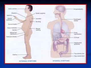
CYANOSIS • BLUISH discoloration of SKIN & MM due to doxyhemoglobin. • Desaturated arterial blood Central cyanosis. • Peripheral cyanosis Normal Art. Sat. • Detected; clinically ( sat below 85% ), pulse oximetry, or ABG.
CAUSES OF CYANOSIS • Central cyanosis: • Respiratory • CNS • Muscular • Cardiac • Peripheral cyanosis: • CHF • Shock • Acrocyanosis • Abnormal hemoglobin
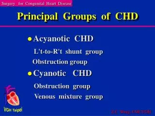
Types of Cyanotic CHD • With pulmonary blood flow; • D-TGA • Truncus arteriosus • TAPVD • DORV • Single ventricle no ps. • With pulmonary blood flow; • TOF • Critical pulmonary stenosis • Pulmonary Atresia
D-TGA • Parallel circulation not in series • Body----RA----RV----AO----Body • Lungs---LA----LV----PA----Lungs • Poor mixing • Hypoxia & Acidemia • Hyperventilation • Increased pulmonary flow • CHF • Myocardial depression
TRUNCUS ARTERIOSUS • Complete mixing so cyanosis is minimal • VSD always present • Identical pressures in both ventricles • Systemic saturation is proportional to pulmonary blood flow ( PBF ) • PVR & PA.s caliber determine PBF • CHF is a usual presentation • Eisenmenger syndrome may develop • AS & AI are complicating factors
TAPVR • NON-Obstructed TAPVR • Similar hemodynamics to a large ASD • Lt to Rt shunt magnitude is determined by RV compliance & ASD size • Rt heart & pulmonary volume overload • Complete mixing at RA level • Minimal cyanosis due to large PBF • Slight PA pressure elevation
TAPVR, CONT; • Obstructed TAPVR • Pulmonary venous hypertension & secondary PA & RV hypertension • Less RV & PA volume overload • Pulmonary venous oedema • More cyanosis & respiratory distress • Complete mixing
TRICUSPID ATRESIA • Increased RA pressure & size • Dependant on the ASD or PFO • Dilated LA & LV • Complete mixing in LV • With TGA PBF & SAT. • Variable degree of cyanosis
EBSTEIN ANOMALY • Variable degrees of cyanosis depending on amount of Rt to Lt shunt across ASD or PFO • Hugely dilated RA • Incompetent TV • Atrialized & small poorly functional RV • RVOTO & PS can be associations • SVT ( WPW ) can be associated
TETRALOGY OF FALLOT • Decreased PBF & Amount of RT to LT shunt determine degree of cyanosis • PBF is mainly determined by RVOTO & PS • Pink TOF = mod RVOTO & balanced shunting across the VSD • Increased PBF can result from PDA or MAPCAS • Equal ventricular pressures
PULMONARY ATRESIA • With VSD • An extreme form of TOF • Early cyanosis when PDA closes • Ductal dependant • CHF with MAPCAS / PDA • Balanced form
PULMONARY ATRESIA CONT; • Without VSD • Early marked cyanosis ( extremely decreased PBF ) • Ductal dependant • RV sinusoids • RV decompression by TR • ASD or PFO mandatory
SUMMARY • Central cyanosis is the hallmark of cyanotic CHD • Cyanosis is a serious symptom or sign • Variable degrees of cyanosis occur due to variability in PBF, PVR, RVOTO, presence & size of shunting site e.g. ASD and hemoglobin concentration • CHF can occur in cyanotic CHD due to increased PBF • Pulmonary AVMs & Methemoglobinemia are non-cardiac causes of central cyanosis.