Download
1 / 53
560 likes | 1.13k Views
Skin and Body Membranes. Chapter 4. What are body membranes?. Cover surfaces, line cavities, protect organs 2 groups Epithelial membranes – cutaneous, mucous, serous Connective Tissue membranes – synovial membranes. Epithelial Membranes. Coving and lining membranes

E N D
Skin and Body Membranes Chapter 4
What are body membranes? • Cover surfaces, line cavities, protect organs • 2 groups • Epithelial membranes – cutaneous, mucous, serous • Connective Tissue membranes – synovial membranes
Epithelial Membranes • Coving and lining membranes • Do contain epithelial sheet, but all have connective tissue underneath • Simple organs
Epithelial Membrane - Cutaneous • Skin • Superficial – keratinizing stratified squamous epithelium • Deep (dermis) – dense connective tissue • Exposed to air and dry membrane Figure 4.1a
Epithelial Membrane - Mucous • Epithelium – varies per site, most often stratified squamous or simple columnar • Loose connective tissue – called lamina propria • Lines all body cavities open to exterior • What? • Respiratory, Digestive, Urinary, Reproductive • Wet membranes, bathed in secretions (or urine) • Adopted for absorption or secretion • Almost all produce mucus but urinary does not
Epithelial Membranes - Serous • Superficial - Simple squamous • Deep – loose connective areolar • Line cavities closed to exterior (except dorsal body cavity and joint cavities) • What? Figure 4.1c–d
Epithelial Membranes - Serous • Come in pairs • Parietal layer – lines wall of ventral body cavity, folds over on itself to form, always connected to wall • Visceral layer – covers outside layer of organ • Compare to balloon • Two layers separated by serous fluid – secreted by both layers, lay very close together • Why? • Name of serous membrane based on location • Peritoneum • Pleura • Pericardium
Connective Tissue MembranesSynovial Membranes • Areolar connective tissue • Line fibrous capsules surrounding joints • Smooth surface • Secrete lubrication • Lines sacs of connective tissue of bursae and tendon sheaths Figure 4.2
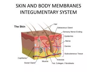
The Integumentary SystemSkin • Cutaneous Membrane • Include skin, sweat and oil glands, hair, and nails • Mostly protective
Functions Table 4.1
Functions • Upper layer contains keratin • Cornified (hardened) – protect from water loss • Rich capillary network and sweat glands • Controlled by nervous system • Regulate heat loss • Mini-excretory system • Urea, water, and salts lost • Manufac. Proteins imp. to immunity and Vit. D synthesis • Cutaneous Sensory Receptors in skin – touch, pressure, temperature, pain (part of nervous)
Structure of the Skin • Epidermis – outermost layer • Stratified squamous able to keratinize • Dermis – underlying tissue • Dense connective tissue • Firmly connected, friction can separate • Blister – What happens? • Subcutaneous layer or hypodermis – adipose tissue • Not skin, anchors to organs • Shock absorber, insulator • Curves of woman
Epidermis • Up to five layers thick • You get to learn them all in 2A!! • Avascular – shaving! • Most cells keratinocytes • Bottom layer stratum basale anchored to dermis by epidermal cells, receive nutrients via diffusion from dermis, millions of new cells daily, pushed outward (DEAD) • You will shed 40lb of skin in lifetime Figure 4.3
Melanin • Pigment – yellow to brown to black • Produced by melanocytes found in stratum basale • Exposed to sun, produce more melanin • Collected in melanosomes, taken up by keratinocytes • Act as umbrella protecting DNA • Freckles or moles are concentrated melanin
Herpes Simplex and the sun! • Sun exposure damages skin • Depresses immune system • Develop cold sore more often after sun bathing • Overexposure of skin to UV rad. can lead to skin cancer • Black individuals seldom have skin cancer • Power of melanin
Dermis • Strong, stretchy envelop that holds you together • Hide – leather goods treated dermis • Dense Connective Tissue • Papillary layer – upper • Reticular layer - lower • Varies in thickness – feet/hands compared to eyelids Figure 4.4
Papillary Layer of Dermis • Uneven, projections called dermal papillae • Contain capillary loops which nourish epidermis • Some contain pain and touch receptors • Hands and feet – looped and whorl patterns for friction and grip • Determined genetically - fingerprints Figure 4.4
Reticular Layer of Dermis • Blood vessels, sweat and oil glands, deep pressure receptors • Phagocytes prevent bacteria
Dermis • Collagen and elastic fibers • Collagen – toughness, attract and bind water, keep skin hydrated • Elastic – give skin elasticity • As age both decrease, subcutaneous looses fat, sag and wrinkle • Blood vessels to regulated heat • Hot – hot blood, radiate heat • Cold – bypass capillaries • Rich nerve supply – chapter 7
Decubitis Ulcers • Restriction of blood supply results in cell death, possibly skin ulcers • Bed sores – bedridden patients not turned, pressure points • Permanent damage results in ulceration Figure 4.5
Skin Color • 3 pigments contribute to skin color • Melanin – amount and kind in epidermis • Carotene – deposit in stratum corneum and subcutaneous tissue, orange-yellow pigment • Oxygen-rich hemoglobin – in dermal blood vessels • Think about variations!
Cyanosis • Hemoglobin poorly oxygenated, blood and skin of Caucasians appear blue • Heart failure, breathing disorders • In black people, skin does not appear blue due to masking melanin – mucous membranes and nails http://meded.ucsd.edu/clinicalimg/upper_cyanosis.htm
Emotions and Skin Color • Redness (erythema) – embarrassment, fever, hypertension, inflammation, or allergy • Pallor (blanching) – emotional stress (fear, anger), anemia, hypotension, impaired blood flow • Jaundice (yellow) – liver disorder excess bile pigments absorbed by blood, transported and deposited in tissues • Bruises – escaped blood has clotted in tissue, hematomas, deficiency Vit C or hemophilia
Appendages of the Skin • Cutaneous Glands • Hair and hair follicles • Nails • Arise from epidermis, play role in homeostasis
Cutaneous Glands • All exocrine glands – release secretions to skin surface • Formed by cells of stratum basale, push into dermis • 2 Types • Sebaceous (oil) glands • Sweat glands
Cutaneous GlandsSebaceous Glands • All over body except hands and feet • Most ducts empty into hair follicle, some to surface of skin • Sebum = product • Oily substances • Fragmented cells • Lubricant – soft, moist, hair not brittle • Chemicals to kill bacteria • Become very active when male hormones produced = oily skin Figure 4.6a
Imbalance of Sebaceous Glands • Whitehead – gland blocked by sebum • Blackhead – material oxidizes and dries • Acne – active infection of glands • Mild or severe, perm. scaring. • Seborrhea (cradle cap) – overactive sebaceous gland, raised pink lesions form yellow brown crust http://www.medical-look.com/Skin_diseases/Acne.html
Cutaneous GlandsSweat Glands – Sudoriferous Glands • Widely distributed in skin - +2.5 mil/person • 2 types • Eccrine glands – more #’s, all over body • Produce sweat • Effective heat regulation • Nerve endings, stimulate sweat production • Apocrine glands – Axillary and genital areas • Larger than eccrine glands • Ducts empty into hair follicle • Puberty, under direction of androgens (♂ hormones) • Activated by nerves during pain, stress, sexual foreplay
Sweat – Eccrine Gland • Clear secretion • Mostly water + salts (NaCl), Vit C, metabolic waste (urea), lactic acid • Acidic – pH 4-6, inhibits bacterial growth • Reaches surface through pore • Pores on face not sweat pores, but external outlets of hair follicle • Excreted when temp. ↑, can lose up to 7 L of water Figure 4.6b
Product of Apocrine Gland • Fatty acids and proteins in addition to eccrine secretion • Milky or yellowish color • Odorless unless bacteria on skin use proteins for growth – musky, unpleasant odor • Produced almost continually http://academic.kellogg.cc.mi.us/herbrandsonc/bio201_McKinley/Skin.htm
Hair and Hair FolliclesHair • Has lost most of evolutionary purpose • Some protective features – head, eyes, nose
Hair Figure 4.7a • Produced by hair follicle • Flexible epithelial structure • Shaft – projection out of skin • Hair formed from well nourished stratum basale epithelial in matrix (growing) • As push farther from growing region, keratinize and die • Most of hair shaft dead cells like epidermis Figure 4.7c
Hair Figure 4.7b • Medulla, Cortex, Cuticle • Cuticle – single layer of cells that overlap, shingles • keeps hair apart, matting • heavily keratinized – strength, inner compacted • Wears out at ends – splint ends • Melanocytes – hair bulb, produce hair color, melanin – yellow, rust, brown, black Figure 4.8
More Hair • Different shapes and sizes – eyebrows, head, body, etc. • Oval hair shaft – smooth, wavy • Flat, ribbonlike – curly or kinky • Round shaft – straight and course • Found all over body except hands, feet, lips, nipples • Among fastest growing tissue • Hormones account for development of hairy regions – scalp, public, axillary
Hair Follicle • Epidermal sheath – inner, epithelial tissue, forms hair • Dermal sheath – outer, dermal connective tissue, supplies blood to ES and supports • Papilla provides blood supply to hair bulb
Hair Follicle • Hair follicle slanted • Arrector pili muscle • Connects hair follicle to dermal tissue • Muscles contract, hair stands, dimpling of skin • Keeps animals warm in winter, added layer of warmed air • Not useful to humans
Nails • Modification of epidermis, like hoof or claw • Free edge, body, root • Nail fold or cuticle • Stratum basale – nail bed, thickened proximal area is nail matrix (growth), cells heavily keratinized and dead • Colorless, look pink due to blood supply • Lunula – white crescent Figure 4.9
Homeostatic Imbalances of Skin • When something goes wrong, very visible • Most diseases caused by allergies, bacterial, viral, or fungal infections • More damaging are burn and cancers
Infections and AllergiesAthlete’s Foot • Itchy, red, peeling skin • Fungal infection • Tinea pedis • Can affect other areas of skin • Ringworm http://www.squidoo.com/natural-cure-for-athletes-foot-x
Infections and AllergiesBoils and Carbuncles http://www.mayoclinic.com/health/medical/IM01272 • Inflammation of hair follicles and sebaceous glands • Carbuncles – boils caused by bacterial infections like Staphylococcus aureus http://www.dshs.state.tx.us/idcu/health/antibiotic_resistance/mrsa/picpage.asp
Infections and AllergiesCold Sores • Fluid filled blisters • Herpes simplex virus • Localized to cutaneous nerve where dormant until stress, fever, UV radiation • Lips or oral mucosa http://www.stanford.edu/group/virus/herpes/2000/herpes2000v1.html
Infections and AllergiesContact Dermatitis • Itching, redness, swelling, eventual blistering • Exposure to chemicals that elicit allergic rxn • Poison ivy http://mmcenters.discoveryhospital.com/shared/enc/img_htm/Derm-12.htm
Infections and AllergiesImpetigo • Pink, water filled, raised lesions • Normally around nose and mouth • Develop yellow crust than rupture • Caused by staphylococcus infection • Common in elementary school aged children http://www.impetigodoctor.com/impetigo-photo.htm
Infections and AllergiesPsoriasis • Chronic condition • Overproduction of skin cells • Reddened epidermal lesions covered with dry, silvery scales • Itch, burn, crack, bleed • Autoimmune disorder • Trauma, stress, hormonal changes, infections http://www.psoriasite.com/
Importance of skin • Skin only paper towel thick, when damaged affects all organs systems • Metabolism accelerates or impaired • Immune system changes • Cardiovascular system may fail
Burns • Burn – tissue damage and cell death by intense heat, electricity, UV radiation, chemical • 2 life threatening problems result from burns • 1) Loss of supply of fluids containing proteins and electrolytes that seep from burns • Dehydration and electrolyte imbalance follow – shut down of kidneys and circulatory shock • 2) Infections – leading cause of death • Burned skin sterile for 24 hours • Pathogens – bacteria and fungus multiply in nutrient rich dead tissue • Immune system suppressed
Burns - Classification • First degree burns – only epidermis damaged, red, swollen, sunburn • Second degree burns – epidermis and upper region of dermis, red, painful, blisters, no permanent scars if prevent infection • Both 1 and 2 partial-thickness burns • Third degree burns – full thickness burn, destroy entire thickness of skin • Appears blanched or blackened, nerves destroyed so area not painful • Regeneration not possible, grafting to cover tissue
When are burns critical? • Over 25% of body has second degree burns • Over 10% of body has third degree burns • Third degree burns on face, hands or feet • Face dangerous why?
Skin Cancer • 1 in 5 Americans • Basal Cell Carcinoma • Least malignant and most common • Cell of stratum basale • Do not form keratin, no boundary between dermis and epidermis • Usually on sun exposed area • Slow-growing • 99% cure rate when surgically removed Figure 4.12
Skin Cancer • Squamous Cell Carcinoma • Cells of stratum spinosum • Scaly, reddened papule to shallow ulcer with firm raised border • Scalp, ears, dorsum of hands, lower lip • Grows rapidly, metastasizes to lymph nodes if not removed • Sun induced • Surgical removal and/or radiation therapy