Download
1 / 1
10 likes | 119 Views
The Role and Mechanism of PPAR in the Transcriptional Regulation of its Target Genes. Jinlu Cai 1 , Henry L. Keen 2 ,Thomas L. Casavant 3,4,5 , and Curt D. Sigmund 2

E N D
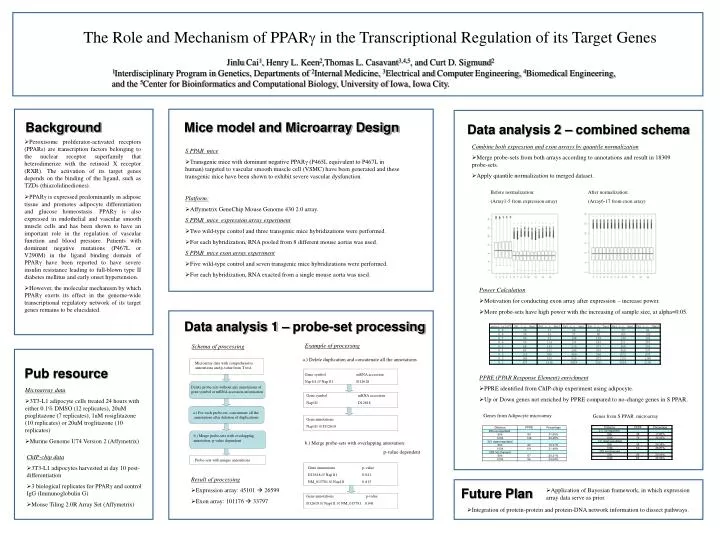
The Role and Mechanism of PPAR in the Transcriptional Regulation of its Target Genes Jinlu Cai1, Henry L. Keen2,Thomas L. Casavant3,4,5, and Curt D. Sigmund2 1Interdisciplinary Program in Genetics, Departments of 2Internal Medicine, 3Electrical and Computer Engineering, 4Biomedical Engineering, and the 5Center for Bioinformatics and Computational Biology, University of Iowa, Iowa City. Background Mice model and Microarray Design Data analysis 2 – combined schema • Peroxisome proliferator-activated receptors (PPARs) are transcription factors belonging to the nuclear receptor superfamily that heterodimerize with the retinoid X receptor (RXR). The activation of its target genes depends on the binding of the ligand, such as TZDs (thiazolidinediones). • PPARγ is expressed predominantly in adipose tissue and promotes adipocyte differentiation and glucose homeostasis. PPARγ is also expressed in endothelial and vascular smooth muscle cells and has been shown to have an important role in the regulation of vascular function and blood pressure. Patients with dominant negative mutations (P467L or V290M) in the ligand binding domain of PPARγ have been reported to have severe insulin resistance leading to full-blown type II diabetes mellitus and early onset hypertension. • However, the molecular mechanism by which PPARγ exerts its effect in the genome-wide transcriptional regulatory network of its target genes remains to be elucidated. • Combine both expression and exon arrays by quantile normalization • Merge probe-sets from both arrays according to annotations and result in 18309 probe-sets. • Apply quantile normalization to merged dataset. • S PPAR mice • Transgenic mice with dominant negative PPARγ (P465L equivalent to P467L in human) targeted to vascular smooth muscle cell (VSMC) have been generated and these transgenic mice have been shown to exhibit severe vascular dysfunction. Platform: • Affymetrix GeneChip Mouse Genome 430 2.0 array. S PPAR mice expression array experiment • Two wild-type control and three transgenic mice hybridizations were performed. • For each hybridization, RNA pooled from 8 different mouse aortas was used. S PPAR mice exon array experiment • Five wild-type control and seven transgenic mice hybridizations were performed. • For each hybridization, RNA exacted from a single mouse aorta was used. Before normalization: (Array1-5 from expression array) After normalization: (Array6-17 from exon array) • Power Calculation • Motivation for conducting exon array after expression – increase power. • More probe-sets have high power with the increasing of sample size, at alpha=0.05. Data analysis 1 – probe-set processing Example of processing Schema of processing a.) Delete duplication and concatenate all the annotations Microarray data with comprehensive annotations and p-value from T test. Pub resource Gene symbol mRNA accession Nap1l1 /// Nap1l1 D12618 • PPRE (PPAR Response Element) enrichment • PPRE identified from ChIP-chip experiment using adipocyte. • Up or Down genes not enriched by PPRE compared to no-change genes in S PPAR. Delete probe-sets without any annotations of gene symbol or mRNA accession information • Microarray data • 3T3-L1 adipocyte cells treated 24 hours with either 0.1% DMSO (12 replicates), 20uM pioglitazone (7 replicates), 1uM rosiglitazone (10 replicates) or 20uM troglitazone (10 replicates) • Murine Genome U74 Version 2 (Affymetrix) Gene symbol mRNA accession Nap1l1 D12618 a.) For each probe-set, concatenate all the annotations after deletion of duplications Genes from Adipocyte microarray Genes from S PPAR microarray Gene annotations Nap1l1 /// D12618 • b.) Merge probe-sets with overlapping annotation: p-value dependent b.) Merge probe-sets with overlapping annotation: p-value dependent • ChIP-chip data • 3T3-L1 adipocytes harvested at day 10 post-differentiation • 3 biological replicates for PPARγ and control IgG (Immunoglobulin G) • Mouse Tiling 2.0R Array Set (Affymetrix) Probe-sets with unique annotations Gene annotations p-value D12618 /// Nap1l1 0.041 NM_015781 /// Nap1l1 0.813 • Result of processing • Expression array: 45101 26599 • Exon array: 101176 33797 Future Plan • Application of Bayesian framework, in which expression array data serve as prior. Gene annotations p-value D12618 /// Nap1l1 /// NM_015781 0.041 • Integration of protein-protein and protein-DNA network information to dissect pathways.


![[VI]. Post-Transcriptional Processing and Post-Transcriptional Control of Gene Expression](https://cdn3.slideserve.com/6614804/vi-post-transcriptional-processing-and-post-transcriptional-control-of-gene-expression-dt.jpg)