Download

1 / 21
310 likes | 856 Views
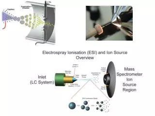
ToF -SIMS – Time of Flight-Secondary Ion Mass Spectroscopy. A surface analytical technique. Routine analytical technique Detailed chemical structure information High sensitivity New primary ion sources (Au, Bi, & buckministerfullerene. General Schematic.

E N D
ToF-SIMS – Time of Flight-Secondary Ion Mass Spectroscopy A surface analytical technique
Routine analytical technique • Detailed chemical structure • information • High sensitivity • New primary ion sources (Au, • Bi, & buckministerfullerene
Principles of ToF-SIMS • Pulsed primary ion beam • Emission of particles – SECONDARY IONS • Ions are mass analyzed by FLIGHT TIMES • Two modes of analysis: static & spectroscopic
TOF-SIMS • Bombard surface with gallium and run through mass spectrometer • Gives both chemical composition of surface and “SEM-like” image of where chemicals are located • Lightly used on lignocellulosic materials • Find concentrations as low as 10 ppm http://www.phi.com/genf.asp?ID=83
Static Mode • Delicate organics (biomaterials) • Undamaged (opposed to X-ray fluorescence microscopy) • Surface sensitive (outermost couple of nm)
Spectroscopic Mode • ONLY mass spectral (MS) data provided • Chemical imaging is POSSIBLE • Raster a micro-focused ion beam (sound familiar???) over surface • Collect MS • Map distribution of species
Organic imaging technique • Previously, limted by most significant signals – polyatomic clusters • For example, most biomaterials dominated by fragments (CxHy+/-) at low mass (< m/z 100) – MORE than one species • NOT DIAGNOSTIC
Higher-order chemical imaging • Larger masses (m/z > 200) more structurally assignable and unique • Chemical mapping possible • Ga+ bombardment doesn’t allow for sufficient sensitivity for good imaging • Polyatomic primary ion sources overcomes deficiency (Aun+, Bin+, C60+) 100x increases in secondary ion yields
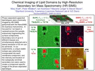
Chemical imaging of pharmaceuticals • Drug-loaded particles can be visualized with Bi3+, whereas with Ga+ they cannot • Due to low intensity of molecular ion peak • Tablet formulations can be studied – distribution of drug, excipient(s), lubricant(s) on surface and in bulk • Thickness & uniformity can be assessed
ToF-SIMS Images 10 micron diameter hair fibers Distribution of materials Nylon mesh – 10 micron depth Plasma cleaned scalpel blade
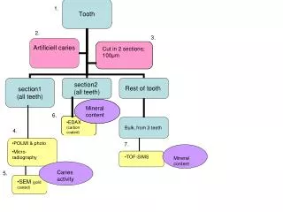
Lignocellulosic biomaterials • The work of Thompson with superoxide (Potassium superoxide) in DMSO found attack in amorphous regions first • Hemicellulose and lignin removed more rapidly than cellulose • The work of Kim with periodate oxidation suggests that the attack on crystalline cellulose proceeds highly heterogeneously • Once an area is damaged, however, the area becomes more susceptible to damage due to loss of crystalline order Thompson, N.S., Corbett, H.M, “The effect of potassium superoxide on cellulose”, TAPPI, 68:12, pp. 68-72, 1985. Kim, U., Kuga, S., Wada, M., Okano, T., Kondo, T. “Periodate oxidation of crystalline cellulose”, Biomacromolecules, 1:488-492, 2000.
TOF-SIMS hypotheses • Lind studied the ability of hydroxyl radicals to induce viscosity loss in cellulose fibers • In their work they found that the decrease in viscosity was proportional to the imparted irradiation dose • This can be read to mean that as the number of hydroxyl radicals increases, so does the cellulose degradation • The work of Lind studied the role of hydroxyl radicals in viscosity loss using ionizing radiation • In their work they found that almost no amount of radical scavenger could protect against depolymerization of the cellulose • This means that hydroxyl radicals produced outside the cellulose surfaces have a minimal effect on degradation and are more likely produced very near to their consumption point Lind, J., Merényi, G., “Hydroxyl radical induced viscosity loss in cellulose fibers”, J. Wood Chem. Technol., 17,(1,2), pp. 111-117 (1997).
TOF-SIMS hypotheses, continued • Metal-induced peroxide cellulose degradation causes the creation of carboxylic acid content • Work of Lind shows that hydroxyl radicals are formed and react very near to the carbohydrate surface • Work of Kleen has been used to measure metals on fiber surface as compared to bulk during bleaching, Found majority of metals, 5 to 55 times bulk, on surface • Not likely to be precipitates due to the fact that the sheets were made at a pH of 5 M. Kleen, Sixth European Workshop on Lignocellulosics and Pulp, 41-44 (2000)
Central hypothesis of work Hypothesis: We are seeing metals bound to carboxylic acid groups caused by radical degradation of cellulose • Metal distribution begins rather homogeneous in the unbleached case, but becomes heterogeneous in the bleached cases • Attack appears heterogeneous concentrated and surface orientated due to the fact it does not appear “deep” enough to be seen by SEM under comparable resolutions Total Ion Image Unbleached Mg Bleached Mg Bleached + 50ppm iron Fe
ESCA • Used to identify carbon and oxygen and the oxidation level • Has been utilized for the detection of carboxyl content (mostly fiber modification work) in literature and is comparable to other methods • We appear to have a difference between the bleached and unbleached samples in COOH content
Viscosity and physical testing • As the load bearing structure of a fiber, the cellulose chains, are being cleaved or “peeled” the mechanical strength of a fiber should decrease • This change should manifest itself in test to include the zero-span tensile and the standard tensile test
Viscosity and physical testing • Chain scission count increases as degradation conditions become more favorable • Zero-span tensile tests show strength decrease as well. Perhaps we also see chemical refining
Summary • TOF-SIMS appears to visualize degradation through the indirect measurement of COOH groups • The analysis of this degradation can be coupled with other techniques including ESCA, viscosity, and zero-span tensile • Degradation appears to be a heterogeneous surface phenonemon
Other work Phytic Acid Chelation • Relatively unstudied chelant that is a product of unwanted by-products of corn • Current data shows performance on par with DTPA/EDTA, but effectiveness is very pulp dependent • Agriculture literature says excellent chelant for iron • Studying it as a bleaching additive and chelant on many different pulp samples and at differing pH