Download
1 / 1

E N D
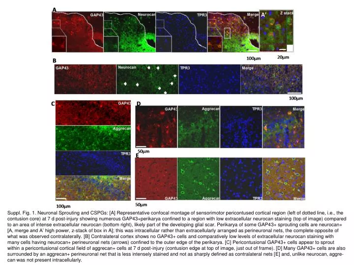
Suppl. Fig. 1. Neuronal Sprouting and CSPGs: [A] Representative confocal montage of sensorimotor pericontused cortical region (left of dotted line, i.e., the contusion core) at 7 d post-injury showing numerous GAP43+perikarya confined to a region with low extracellular neurocan staining (top of image) compared to an area of intense extracellular neurocan (bottom right), likely part of the developing glial scar. Perikarya of some GAP43+ sprouting cells are neurocan+ [A, merge and A’ high power, z-stack of box in A]; this was intracellular rather than extracellularly arranged as perineuronal nets, the complete opposite of what was observed contralaterally. [B] Contralateral cortex shows no GAP43+ cells and comparatively low levels of extracellular neurocan staining with many cells having neurocan+ perineuronal nets (arrows) confined to the outer edge of the perikarya. [C] Pericontusional GAP43+ cells appear to sprout within a pericontusional cortical field of aggrecan+ cells at 7 d post-injury (contusion edge at top of image, just out of frame). [D] Many GAP43+ cells are also surrounded by an aggrecan+ perineuronal net that is less intensely stained and not as sharply defined as contralateral nets [E] and, unlike neurocan, aggre- can was not present intracellularly.