Download
1 / 14
170 likes | 554 Views
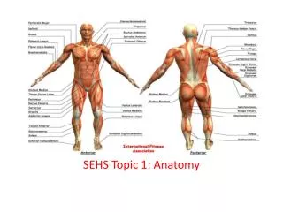
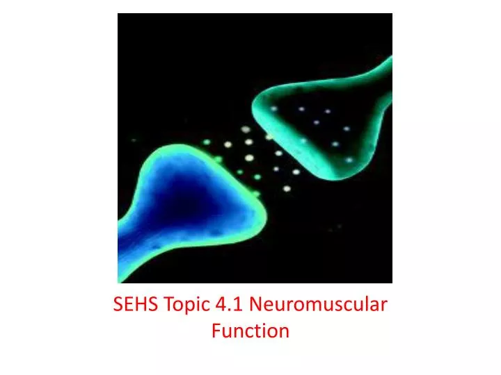
SEHS Topic 4.1 Neuromuscular Function. Label a diagram of a motor unit. Foundation required: A motor unit is made up of a motor neuron and all of the skeletal muscle fibers innervated by that axon. What’s neuron? It’s the functional unit of the nervous system (a.k.a. a nerve cell).

E N D
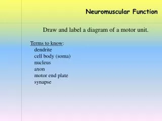
Label a diagram of a motor unit • Foundation required: • A motor unit is made up of a motor neuron and all of the skeletal muscle fibers innervated by that axon. • What’s neuron? • It’s the functional unit of the nervous system (a.k.a. a nerve cell)
Cont’d • http://www.getbodysmart.com/ap/muscletissue/nervesupply/motorunit/tutorial.html • The large and complex terminal formation by which the axon of a motor neuron establishes synaptic contact with a striated muscle fiber
Cont’d • Label this: use the following word bank: tranverse(t) tubule; axon; synaptic vesicle containing neurotransmitters; synapse; sarcolemma; motor end plate; sarcoplasmic reticulum; neuromuscular junction
Explain the roles of neurotransmitters (NTs) in stimulating muscle contraction • What are neurotransmitters (NTs)? • Endogenous chemicals that transmit signals from a neuron to a target cell across a synapse. • They are packaged into synaptic vesicles clustered beneath the membrane in the axon terminal, on the presynaptic side of a synapse. • They are released into and diffuse across the synapse (a.k.a. synaptic cleft), where they bind to specific receptors in the membrane on the postsynaptic side of the synapse
Cont’d • The key NTs for skeletal muscle contraction include” • Acetylcholine (ACh) and cholinesterase • In the peripheral nervous system, acetylcholine activates muscles, and is a major neurotransmitter in the autonomic nervous system. • When acetylcholine binds to acetylcholine receptors on skeletal muscle fibers, it opens gated sodium channels in the cell membrane. • Sodium ions then enter the muscle cell, initiating a sequence of steps that finally produce muscle contraction.
Cont’d • So what happens after a motor neuron has been stimulated by Ach? • In biochemistry, cholinesterase is a family of enzymes that catalyze the hydrolysis of the neurotransmitter acetylcholine into choline and acetic acid, a reaction necessary to allow a cholinergic neuron to return to its resting state after activation
Cont’d: it essentially terminates synaptic trasmission • It is also know as a: degradation enzyme
Explain how skeletal muscle contracts by the SLIDING FILAMENT THEORY • Foundation 1: • Muscle structure (review in part):
Animations • http://www.youtube.com/watch?v=EdHzKYDxrKc • http://www.liquidjigsaw.com/science/animation/animations/bigpicture/muscle-contraction.html
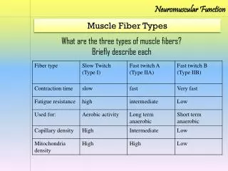
Explain how slow and fast twitch muscle fibers differ in structure and function • Most of us have a 50%-50% make-up of slow vs. fast twitch muscle fibers. Some of their functions and features are shown below and on the next slide.
Cont’d – A more inclusive table • Exercise/sports types: ____________ ______________ ________________