Download
1 / 30
430 likes | 1.28k Views
Complete Blood Count and Anemia. Clinical Pathology. Blood Composition. Separates into three components: Red Blood Cells (RBC’s) White Blood Cells and platelets (buffy coat) Plasma Bottom 1/3 to ½ of tube contains the heaviest of cellular material (the RBC’s).

E N D
Complete Blood Count and Anemia Clinical Pathology
Blood Composition • Separates into three components: • Red Blood Cells (RBC’s) • White Blood Cells and platelets (buffy coat) • Plasma • Bottom 1/3 to ½ of tube contains the heaviest of cellular material (the RBC’s).
Hematocrit=PCV (Packed Cell Volume) • To determine hematocrit, whole blood is centrifuged to pellet the red blood cells. • Plasma remains on the top of the red cells. • The fraction of blood that is packed is the hematocrit and is read as a percentage.
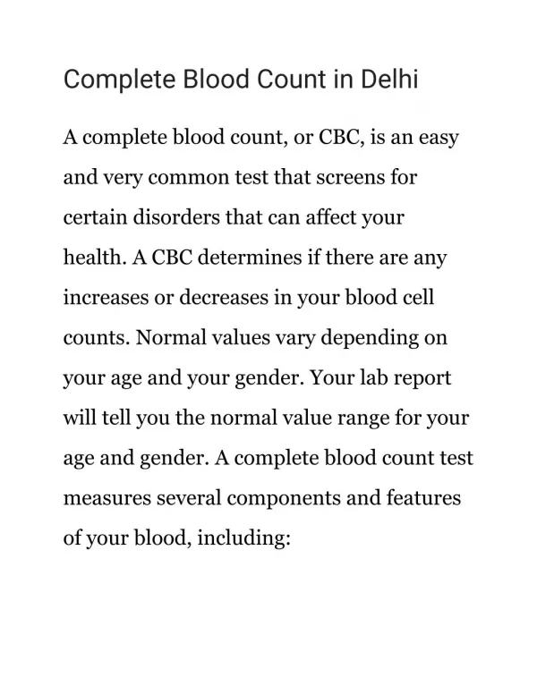
Complete Blood Count • Provides a minimum set of values and is cost effective. • Can be done manually or with automated systems. • CBC should contain: • Packed Cell Volume (PCV or Hct) • Plasma Protein Concentration • Total White Blood Cell count • Blood smear with morphology • WBC differential count • Reticulocyte count
Manual Procedures • PCV- whole blood is collected in anticoagulant, placed in capillary tube, sealed, centrifuged and read. • Total protein- plasma is read with a refractometer.
More Manual Procedures • Absolute WBC: Total number of white blood cells in the blood. • Unopette hematocytometer test kits are used to lyses RBC’s and to make a 1:100 dilution. • WBC’s are counted within the grid and calculated to reflect the WBC in the blood.
Manual Procedures Continued • Differential Leukocyte Count: a relative count is performed by counting and classifying at least 100 leukoctyes. • This gives a percentage of each cell type which is then used to calculate the absolute numbers of each cell type. • May use a counter in order to perform this count.
Instrumentation • Electronic cell counters: based on the principle that cells are poor electrical conductors. • Measured volume of diluted blood is drawn between two electrodes, causing a resistance in the electrical current. • QBC: Quantitative Buffy Coat System • Utilizes differential centrifugation and quantification of cellular elements in a specialized microhematocrit tube.
Red Blood Cell Indices • PCV (hematocrit) • Hemoglobin Concentration • Total red blood cell count • These are used to classify the type of anemia.
Anemia • Literally means “no blood” but clinically means low total blood hemoglobin. • Absolute anemia: most common, caused by failure to produce adequate numbers of cells or by a loss of cells at a rate greater than can be produced.
Clinical Signs of Anemia • Pale mucous membranes • Exercise intolerance • Tachycardia • Panting • Icterus if anemia is caused by RBC breakdown in bloodstream.
Classification of Anemia • By RBC size (MCV): • Macrocytic • Erythrocytes are larger than normal. • Usually in the presence of regenerative anemia. • May be seen in FeLv • May see anisocytosis • Normocytic • Microcytic • Cells are smaller than normal which has been determined by Mean Cell Volume (MCV). • Usually occurs with iron deficiency caused by chronic blood loss or parasitism • By Hemoglobin concentration (MCHC) • Hypochromatic • RBC’s have decreased density of the characteristic hemoglobin color. • Frequently observed in iron deficiency caused by chronic blood loss or parasitism. • Normochromatic
MCV • Describes cells as normocytic, microcytic, or macrocytic. Calculates the average volume of rbc’s. • MCV=(Hematocrit x 10)/RBC count in millions • Ex: • Canine patient with hematocrit of 42% and RBC count of 6 million/ul. • Normal: 66-77
MCV causes of Increases • Reticulocytosis • Congenital issues (poodles) • Cats with FeLv • RBC agglutination • B12 deficiency (rare)
MCV causes of decreases • Abnormal Hgb synthesis (iron deficiency from chronic blood loss is the most common). • Immature animals • Dogs with PSS. • Congenital (Akitas)
MCHC • Mean Corpuscular Hemaglobin Concentration describes cells as normochromatic or hypochromatic. • MCHC= (Hgb)/(Hct) x 100 • Ex. • Same patient as before with Hgb content of 14 g/dL • Normal: 31-36%
MCHC causes if high • Intravascular hemolysis • Inaccurate Hgb reading (Heinz bodies, lipemia, etc). • Machine error • True hyperchromasia does not exist.
MCHC causes if low • Small reticulocytes • Iron deficiency.
Classification According to Bone Marrow Response • Regenerative anemia: • Characterized by evidence of increased production and delivery of new erythrocytes into circulation. • Usually suggests an extra bone marrow cause (blood loss, hemolysis, etc.)., • Diagnosis: • Peripheral blood smear. • Will see macrocytosis, polychromasia with Wright’s stain, reticulocytosis with methylene blue stain, may also see increased numbers of nucleated RBC’s
Nonregenerative anemia: • Indicates anemia is result of bone marrow defect. • No response evident in peripheral blood. • Marrow examination may be helpful with the diagnosis.
Reticulocyte Count • Probably the most important diagnostic tool used in the evaluation of anemia. • Expressed as a % of the RBC’s present. • Corrected to take in account the reduced number of circulating RBC’s in the anemic animal. • Called CRC or Corrected Reticulocyte Count • The lifespan of a normal RBC is about 100 days. • Bone marrow should replace 1 % of the RBC’s daily so the reticulocyte count should be 0.5-1.5%.
Reticulocyte count continued • Expressed as # of retics/100 RBC’s • Some species variation in reticulocyte response exists. • Normal horse and cattle blood do not have reticulocytes. • CRC= (patient Hct)/(Normal Hct) x reticulocyte count
Example • Dog with an observed reticulocyte count of 9 % and Hct of 25%. Normal Hct is 45. • Interpretation A (expressed in %): • Normal • Less than or equal to 1 in dog • Less than or equal to 0.4 in cat • Mild • Dog: 1-4 • Cat: 0.5-2 • Moderate • Dog: 5-10 • Cat: 2-3 • Marked • Dog: greater than 10 • Cat: 3-4
Blood Loss Anemia • Results from excessive hemorrhage although source can be subtle. • Must determine if blood loss is internal or external. • Possible causes: • Trauma • Persistent bleeding lesions • Thrombocytopenia • Coagulopathies • Heavy parasitism • Iatrogenic causes
Acute Blood Loss • Anemia due to loss of blood in a sudden episode. • All RBC parameters are normal for the first 12 hours. • Hypovolemic shock can be apparent prior to a decreased PCV. • Anemia will be normocytic, normochromatic, and apparently unresponsive with a low CRC. • By day 4-5, the retic count increases and the anemia appears responsive.
Chronic Blood Loss • Blood is lost slowly and continuously for a period of time. • Body compensates for anemia by lowering oxygen-hemoglobin affinity, preferential shunting of blood to vital organs, increased cardiac output (tachycardia), and increased levels of erythropoietin. • Anemia remains unresponsive unless iron stores are depleted. • With decreasing iron stores, erythropoiesis is limited and RBC’s become smaller and deficient in Hgb (microcytic and hypochromic). • Clinical signs include lethargy, weakness, decrease exercise tolerance, anorexia, pallor, lack of grooming, mild systolic murmur.
Diagnostic Tests • Hemogram: may see increased WBC and platelets. • Total protein: decreased • Coagulation testing: platelet count, PT, PTT, ACT. • Fecal Float: Hookworms, Whipworms • Fluids analysis from body cavities
Hemolytic Anemias • Result of increased erythrocyte destruction within the body. • Intravascular hemolysis: desctruction of erythrocyctes within the blood vessels and loss of Hgb from the cells. • Extravascular hemolysis: RBC’s are lysed following phagocytosis.
Differentials • Immune-mediated disease: AIHA, drug induced, neonatal isoerythrolysis. • Parasitic: Ehrlychiosis, Babesiosis, Hemobartonellosis, Anaplasmosis. • Toxic: Heinz body anemias, snake venom, bacterial toxins. • Infectious: EIA, Leptospirosis, Clostridia • Fragmentation: Splenic torsion, Splenic neoplasia, DIC