Download
1 / 44
440 likes | 552 Views
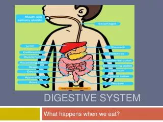
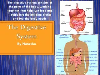
Explore the major structures of the human digestive system including the mouth, esophagus, stomach, small and large intestines. Compare with cat digestive system, highlighting differences such as liver lobes.

E N D
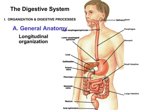
Lab 9 – Digestive System Anatomy Dr. Kim Wilson
Lab Manual Reference • Exercise 38 pg. 575
PART A: ANATOMY OF THE HUMAN DIGESTIVE SYSTEM • INSTRUCTIONS: • Using the human torsos, locate as many as possible of the following structures from the list, "Major Structures of the Human Digestive System." Use your textbook for reference. • Answer related questions on the Questions Sheet.
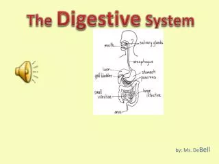
MAIN ORGANS OF THE DIGESTIVE SYSTEM • 1. MOUTH ‑Lips ‑Cheeks ‑Hard Palate ‑Soft Palate ‑Uvula uvula
Pharynx ‑ Fauces The passage from the back of the mouth to the pharynx, bounded by the soft palate, the base of the tongue, and the palatine arches. ‑ Oropharynx ‑ Laryngopharynx
‑Fundus ‑Body ‑Pylorus ‑Lesser Curvature ‑Greater Curvature ‑Cardiac Sphincter ‑Pyloric Sphincter ‑Rugae Stomach Pylorus part of the stomach
Small Intestine ‑ Duodenum ‑ Jejunum ‑ Ileum ‑ Plicae Circulares
Large Intestine • Ileocecal Valve • Cecum • Colon • Ascending Colon • Transverse Colon • Descending Colon • Sigmoid Colon • Hepatic Flexure • Splenic Flexure • Rectum • Anal Canal • Anus • Haustra • Taenia Coli
Ileocecal Valve • Cecum • Colon • - Ascending Colon • - Transverse Colon • - Descending • Colon • Sigmoid Colon • Hepatic Flexure • Splenic Flexure • Rectum • - Anal Canal • - Anus • Haustra • Taenia Coli
ACCESSORY ORGANS OF THE DIGESTIVE SYSTEM SALIVARY GLANDS ‑ Parotid Glands ‑ Submandibular Glands ‑ Sublingual Glands
Root • Tip • Body • Papillae: Vallate, Fungiform, Filiform
Teeth • Crown • Neck • Root • Incisors • Cuspids(Canines) • Bicuspids(Premolars) • Tricuspids(Molars)
Teeth • Crown • Neck • Root • Incisors • Cuspids(Canines) • Bicuspids(Premolars) • Tricuspids(Molars)
Liver LIVER ‑Lobes: Left (1); Right (3) ‑Hepatic Ducts: Right & Left; Common
MEMBRANES OF THE ABDOMINAL CAVITY (PERITONEUM) • Parietal Peritoneum - the part of the peritoneum that lines the abdominal wall 2. Visceral Peritoneum - the part of the peritoneum that lines the abdominal viscera • Mesentery: A layer of connective tissue that is in vertebrates. It supports portions of the small intestine, protects nerves and blood vessels from the central system to the organ. • Transverse Mesocolon • Greater Omentum: A fat-filled, web-like structure draped over the internal organs; it protects the pancreas. • Lesser Omentum: It connects the stomach and liver, and has two portions- the gastrohepatic ligament, and the hepatoduodenal ligament. 6. Falciform Ligament
PART B: ANATOMY OF THE CAT DIGESTIVE SYSTEM • INSTRUCTIONS: • Obtain your assigned cat and locate each of the structures from the list, "Major Structures of the Cat Digestiven System." Use your lab manual (Color Photo Gallery) and the Rust lab manual for reference. • Answer related questions on the Questions Sheet.
Villi You would have to cut open the duodenum to see the villi.
1 Parietal Peritoneum 2 Visceral Peritoneum 3 Small Intestine 4 Colon 5 Greater Omentum 6 Kidneys 7 Right Renal Vein 8 Left Renal Vein 9 Right Renal Artery 10 Left Renal Artery 11 Inferior Mesenteric Artery 12 Left Iliolumbar Artery and Vein 13 Inferior Vena Cava 14 Femoral Artery 15 Femoral Vein 16 Superior Mesenteric Vein 17 Hepatic Portal Vein 18 Gastrosplenic Vein 19 Superior Mesenteric Artery 20 Greater Saphenous Vein 21 Urinary Bladder 22 External Iliac Artery 23 Internal Iliac Artery 24 Pancreas 25 Rectum
Differences in Human and Cat Digestive Systems The cat has six lobes in their liver, and humans only have four lobes.