Download
1 / 81
870 likes | 1.02k Views
HEART FAILURE. Institute of Pathological Physiology Martin Vokurka mvoku@lf1.cuni.cz WS 200 8 /0 9. normal situation. heart failure. congestion. backward. normal situation. heart failure. decreased cardiac output. congestion. forward. backward.

E N D
Institute of Pathological Physiology Martin Vokurka mvoku@lf1.cuni.cz WS 2008/09
normal situation heart failure congestion backward
normal situation heart failure decreased cardiac output congestion forward backward Insufficient organ perfusion – muscles, kidneys, skin, GIT…
heart insufficientlyfilled decreased venous returne.g. shock bad filling of the ventricles(e.g. constrictive pericarditis) decreased cardiac output
heart changes heart failure decreased cardiac output congestion compensatory events
HEART FAILURE pathophysiologic state in which an abnormality of cardiac function is responsible for the failure of the heart to pump blood at a rate commensurate with the requirements of the metabolizing tissues and/or can do so only from an abnormally elevated diastolic volume decrease of cardiac output increase of the ventricular filling pressure (enddiastolic pressure, EDP)
Heart failureis not only failing of the heart as a pump, but it issystemic disorder with activationof hormonal processes, with changed metabolism, changed regulation of water-mineral balance, with cytokines involved, heart changes, changesof gene expression etc. HEMODYNAMIC ASPECTS NEUROHUMORAL ASPECTS CELLULAR AND GENE EXPECT
Frequency of heart failure In the Czech Rep. the prevalence is about 1-2 % (i.e. 100 000 of patients) The number of patients is increasing –among others due to successful treatment of other heart diseases
TYPES OF HEART FAILURE - LEFT-SIDED - RIGHT-SIDED - BOTH-SIDED according to the failing ventricle
MAIN SYMPTOMES 1. CONGESTION - left-sided - DYSPNEA, LUNG EDEMA - right-sided - LOWER EXTREMITY EDEMAS, HEPATOMEGALY… 2. DECREASED CARDIAC OUTPUT WEAKNESS, FATIGUE, DECREASED ORGAN PERFUSION
Causes of heart failure Myocardial failure - defect in myocardial contraction (ischemia, cardiomyopathy) - loss of myocardium (myocardial infarction) Excessive, long-term hemodynamic burden - increased pressure burden (systemic or lung hypertension) - increased volume burden (valvular abnormalities) - hyperkinetic cirkulation (increased CO) Most commonly it is the combination of CHD and arterial hypertension
In heart failure CO decreases Activation of compensatory mechanisms trying to increase CO backto normal values How are the distinct mechanism influencing the COregulated ?
Cardiac output (CO) =heart rate (HR) × stroke volume (SV) 70 /min 70 ml 4 900 ml/min
Heart rate • vegetative nerves • (disturbances in) heart rhythm • has impact of heart cycle duration, mainly shortens diastole – when the heart is filling with blood • Increases CO but high rates decrease the ventricle filling and heart is easier exhausted
Cardiac output (CO) =heart rate (HR) × stroke volume (SV) 70 /min 70 ml 4 900 ml/min
Stroke volume • preload • contractility • - afterload • * How is the heart filled before the systole • * What is its „force“ of contraction • * What is resistance against the pumping
Preload filling of the heart at the end of the diastole enddiastolic volume = EDV Frank-Starling mechanisms Volume in the ventricle corresponds to the pressure – enddiastolic pressure, EDP, filling pressure
Factors influencing preload • Venous return • total blood volume • blood distribution (position of the body, intrathoracic pressure, venous tonus…) • atrial systole • size of ventricle cavity • - intrapericardial pressure • Low preload is the cause of the decreased CO in case of syncope and shock • In heart failure the preload is not decreased but it is increased as one of the the compensatory mechanisms
The relation between the filling of the ventricleand the intraventricular pressure diastolic filling curve volume: EDV - enddiastolic volume pressure: EDP - enddiastolic pressure, filling pressure - amount of the blood in the ventricle - properties of the ventricle wall
J.Kofránek EDP intraventricular pressure ventricle volume
enddiastolicvolume (EDV) enddiastolicpressure (EDP) preload changes in the ventricle geometry pressure “transfer” to the regions „precceding the heart“ dilatationincreased wall tensionincreased oxygen consumptionfailure of Frank-Starling mech. ventricle wall properties(compliance)ischemia - impaired relaxationfibrosishypertrophy/dilatation P L - lung edemaP - e.g. hepatomegaly V
Enddiastolic volume Systolic volume preload Systolic residual volume Intraventricular pressure Isovolumic maxmis Isotonic maxims Diastolic filling Ventricle volume J.Kofránek
J.Kofránek Systolic volume Systolic volume Intraventricular pressure Isovolumic maxims Izotonic maxims Diastolic filling Increased preload... Ventricle volume …increases cardiac output.
J.Kofránek Syst. volume Systolic volume Intraventricular pressure Decreased preload... Ventricle volume …decreases cardiac output.
Stroke volume • preload • contractility • - afterload • * How is the heart filled before the systole • * What is its „force“ of contraction • * What is resistance against the pumping
Contractility Increase:sympatic nerves, catecholamines Decreasedischemia, hypoxia, acidosis, proinflammatory cytokines, some drugs etc. Decreased contractility is often the causative mechanism of heart failure.
catecholamines Systolic volume Systolic volume Intraventricular pressure Isovolumic maxims Shifted isovolumicmaxims Catecholamines increase systolic volume …without increasing preload Ventricle volume J.Kofránek
Stroke volume • preload • contractility • - afterload • * How is the heart filled before the systole • * What is its „force“ of contraction • * What is resistance against the pumping
Afterload • the force against which it contracts, the tension or stress developed in the ventricular wall during ejection • - arterial pressure- systemic vascular resistence- blood viskosity • geometry of the ventricle (Laplace law) • T = P × r / d • Increased volume of the ventricle and thiner wall (i.e. dilatation) increase afterloadcontribute to the decrease of COincrease requirements for oxygen
Increase of „afterload“ Syst. Syst. volume volume Systolic volume Intraventricularpressure Izovolumic maximx Increase of „afterload“ does not chane SV Isotonic maxims Diastolic filling ... but preload is increased Ventricular volume J.Kofránek
Types of heart failure According to the ventricle Acc. to the intensity • left-sided • right-sided (cor pulmonale due to lung diseases, lung embolism etc.) • both-sided According to the course According to the CO • acute • chronic (development of the compensatory mechanisms): • compensated • decompensated • low-output (most) • high-output (hyperkineticcirkulation)
whether the principal abnormality is - the inability to contract normally and expel sufficient blood (systolic failure) - or to relax and fill normally (diastolic failure) Systolic failure Blood ejection from the ventricle is disturbed Stroke volume might be maintaind at the costs of increased EDV (and EDP)
Ejection fraction EF = SV / EDV the ratio of stroke volume to end-diastolic volume normal value = 67 ± 8 percent SV = 70 ml, EDV = 120 ml EF = 70 / 120 = 58 %
Normal heart stimulated by the sympatic nerves - EF increases, SV increases (contractility increased) Heart with noncompensated systolic failure - EF low, SV low Heart with compensated systolic failure and increased preload - EF low, SV might be normal (EDV is increased)
EDV1 End of diastole 1
SV1 ESV1 EF1 = SV1/EDV1 End of systole 1
SV2 ESV2 EF2 = SV2/EDV2 EF2> EF1 End of systole 2
EDV2 End of diastole 2
SV3 ESV3 EF3 = SV3/EDV3 End of systole 3
SV1 ESV1 EF1 = SV1/EDV1 EF1> EF3 SV1 = SV3 End of systole 1
Diastolic failure • usually the decrease in compliance of heart wall • EDP increases • CHD • Hypertension with hypertrophy • Some cardiomyopathies etc.
J.Kofránek filling pressure intraentricular pressure ventricular volume
EDP measurement heart catheterization as a pulmonary wedge pressure
Evaluation / monitoring of hemodynamic heart function • - EF (ultrasound) • - cardiac output (ultrasound or catheterization) • EDP (catheterization) • - Heart rate (HR) • - Blood pressure (BP)
Symptoms of heart failurefrom the hemodynamic point of view Low CO Weakness, fatigue, decreased organ perfusion incl. kidneys, muscles - redistribution of CO FORWARD Blood congestion in organs from which blood is collected to the failing ventricle Edemas etc. BACKWARD
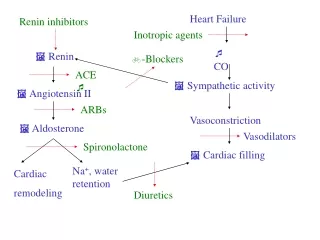
Apart from hemodynamicchanges heart failure is characterized by important involvement of compensatory mechanisms, mainly neurohumoral, which can, however, if persisting, lead to further progression of failure. Another changes involve the heart itself. Compensatory mechanisms – can in short-term have a positive role, in long-termpersistence contribute to theworsening of the failure.
Main compensatory mechanisms in heart failure • They lead to incrase (maintain) CO • Sympatic activity • Increase of preload • Salt and water retention • Myocardium changes • Short-term effective, long-term have deletirious effectsthemselves and contribute to the symptoms and progressionof HF • Vitious circle
Sympatic activity inheart failure • Heart rate • Contractility • Venous return • CO
Negative consequences Tachycardia: Increase in oxygen consumption shortening of the diastole (impairment of diastolic filling and myocardial blood flow) Increased risk for arrhytmias