Download
1 / 36
360 likes | 513 Views
What are we measuring in fMRI?. Caroline Catmur Jack Kelly. In BOLD fMRI, we are measuring: the inhomogeneities introduced into the magnetic field of the scanner… as a result of the changing ratio of oxygenated:deoxygenated blood…

E N D
What are we measuring in fMRI? Caroline Catmur Jack Kelly
In BOLD fMRI, we are measuring: the inhomogeneities introduced into the magnetic field of the scanner… as a result of the changing ratio of oxygenated:deoxygenated blood… via their effect on the rates of dephasing of hydrogen nuclei. Ehhh???
Physics: underlying principles • Hydrogen nuclei (1H): positively charged particles which spin around their axes, producing a (small) magnetic field. • MDM: magnetic dipole moment: vector of the magnetic field of the nucleus. • When placed in a uniform magnetic field, (conventionally indicated by the z axis), the particles’ MDMs align with or against the field. A small percentage more align with the field than against, proportional to the strength of the field, giving the particles a net magnetization.
The MDMs also precess around the axis of the field, at a resonant frequency dependent on the strength of field and type of nucleus, eg 64MHz for 1H in a 1.5T field.
So what goes on in the scanner? • Place the nuclei (ie the brain) in a uniform magnetic field (the scanner). • The next step: apply an RF pulse, frequency equal to frequency of precession of the nuclei, normally at 90° to the magnetic field. This ‘tips’ the MDMs of those nuclei which have this frequency of precession, ie we only ‘tip’ the 1H nuclei. • So, the MDMs of the 1H nuclei are now at 90° to the main field, ie in the x/y plane.
Terminate the RF pulse and the nuclei relax: their MDMs return to the original orientation in the z dimension, and the energy released during relaxation is what is measured by the receiver coil. • Three different relaxation times of interest in MRI: T1, T2 and T2*.
Phase • Before the RF pulse, all the MDMs precess at the same frequency but not in phase. • After they’re tipped, all precess in phase. Can think of it as all MDMs moving together: this produces a strong signal in the x/y plane. • Once the RF pulse ends, begin to dephase: start to cancel each other out and the signal decays.
Two reasons for this dephasing: inhomogeneities in the magnetic field, and ‘spin-spin’ interactions between neighbouring nuclei. • Possible to correct for dephasing due to inhomogeneities in the field by applying another RF pulse at 180° to the initial pulse. Known as a spin-echo sequence.
Back to those relaxation times • T1 relaxation: time course for the MDMs to return to their original (z) orientation. T2 relaxation: time course of the breakdown of the magnetization in the x/y plane due to spin-spin interactions.
T2* relaxation: time course of the breakdown of the magnetization in the x/y plane due to variations in the magnetic field. The T2* processes can be refocused using a 180° spin-echo sequence, though the T2 processes will still remain. • Different tissues have different T1 and T2 relaxation rates. • T1-weighted scan: measure signal at time when relative difference (between tissue types) in amplitudes of MDMs in z dimension is maximum. • T2-weighted scan: measure at time when relative difference in amplitudes of MDMs in x/y plane is maximum. • To get these different scans, change time between RF-pulse and measurement (TE), and between successive RF pulses (TR).
But why do we need to know all this? • BOLD (blood oxygenation level dependent) contrast: measures inhomogeneities in the magnetic field due to changes in the level of oxygen in the blood. So it’s a T2* contrast. • Oxygenated blood contains oxyhaemoglobin: red blood cells with O2 molecule attached. Not magnetic. • Deoxygenated blood: deoxyhaemoglobin: red blood cells without O2. Magnetic.
So if ratio deoxygenated:oxygenated blood is high, increases inhomogeneities in the magnetic field faster breakdown of magnetism in x/y plane (T2* relaxation) decrease in fMRI signal. • If ratio oxygenated:deoxygenated is high, slower T2* relaxation less decrease in signal. • So we can use the change in fMRI signal to infer the relative oxygenation of the blood.
So how do we get the actual information? • Spatial localisation: ‘gradients’. Small magnetic field gradients (eg 30 mT/m) superimposed onto the main static magnetic field. • Remember that the resonant frequency for a nucleus in a magnetic field depends on the field strength. • So, differences in the resonance frequencies encode the positions of the nuclei along the gradient field. • Switching the small gradients on and off is noisy! • Receiving the information: the RF coil both transmits and receives. A volume coil images any part of the brain; a surface coil gives better images, but only for the nearest part of the brain, due to distortions. A phased array coil is a series of surface coils.
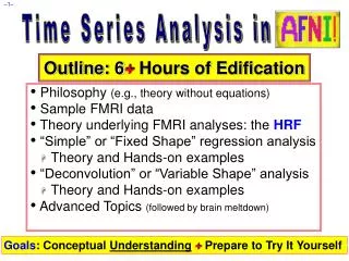
fMRI Outline: • What is BOLD? • Correlation of BOLD with electrophys. • How neurons cause CBF increases • Localising BOLD • Summation of BOLD • Implications for cognitive studies
BOLD and MRI • BOLD = Blood Oxygenation Level Dependent • functional Magnetic Resonance Imaging • Deoxyhemoglobin is paramagnetic and produces a reduced signal, oxyhemoglobin is weakly dimagnetic and doesn’t reduce the signal.
BOLD and electro-physiology: correlation Same area in V1 of cat, Kim DS (2004)
BOLD and electro-physiology: correlation Single unit recording, cat V1, Kim 2004
What causes BOLD? • The purpose of the increase in blood oxygenation is to feed neurons… • …so, what makes a neuron hungry? • (neurons can’t store much energy)
Vascular density • Vascular density is proportional to synaptic density, not soma density
Hungry brains White matter uses ¼ the energy of grey matter per unit volume 62% of mitochondria are in dendrites Attwell & Iadecola 2002
Regulation of blood flow • Is it feedback or feed forward? Activity Uses energy Vascular system must supply more energy Directly commands more blood flow Activity
Regulation of blood flow • Feedforward! Directly commands more blood flow Activity • Energy use does not directly increase blood flow… • …so how does tell CBF to increase?
Monoamines and blood flow • DA, NA and 5HT = vasoconstriction • Cholinergic axons from BF = vasodilation • This complicates neuropsychiatric studies • e.g. schizophrenia, PD, ADHD
Localising fMRI Cat scanner (!); Kim 2004
Correlation of BOLD and single unit Kim 2004
Summation of BOLD BOLD LFP Single unit
Comparing different areas • Different vasculature • Different neuromodulatory control • Different circuitry • BOLD [X] > BOLD [Y] does not mean NEURAL ACTIVITY [X] > ACTIVITY [Y]
What BOLD does not measure • The output of an area • Comparisons of activity between areas • GABA ??????
What does contribute to BOLD • Synaptic activity • Local processing • Sub-threshold neuromodulatory inputs
What does a blob in area X mean? • X has changed its local activity • Change of modulatory inputs arriving at X • Change of inputs arriving at X • (beware: the areas giving rise to the inputs to X may not produce a BOLD signal if their local synaptic activity levels remain constant)
References • Logothetis NK & Wandell BA (2004) Interpreting the BOLD signal Annu. Rev. Physiol 66:735-69 • Attwell D & Iadecola C (2002) The neural basis of functional brain imaging signals Trends in Neurosciences 25:621-625 • Kim DS et al (2004) Spatial relationship between neuronal activity and BOLD functional MRI NeuroImage 21:876-885