Download
1 / 24
260 likes | 542 Views
Human Molecualr Genetics. Luqman Sulaiman MD, MSc, PhD Candidate Molecular & Medical Surgey Karolinska Hospital CMM Chapter One Fri.14-11-08. Aims. Motivate and Encourage Starting point Refresh and boost knowledge How to prepare presentations Have Fun!!!. DNA. Overview

E N D
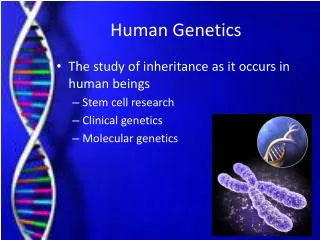
Human Molecualr Genetics Luqman Sulaiman MD, MSc, PhD Candidate Molecular & Medical Surgey Karolinska Hospital CMM Chapter One Fri.14-11-08
Aims • Motivate and Encourage • Starting point • Refresh and boost knowledge • How to prepare presentations • Have Fun!!!
DNA Overview • What is DNA? ( Structure) • Where is located in Eukaryotes? • Types? • Who discovored DNA? when? • Biological Function? • Synthesis? • others
From DNA to Human • 1,2,3- DNA (genes) • 4 -Chromosomes • 5- Tissue , Cells • Human Body
What is DNA? DeoxyriboNucleicAcid (DNA) is a polymer, Nucleoside and Nucleotide are Used interchangebly Nucleotides Nucleoside Base Pairs (Nitrogen bases: Purine and Pyrmaidine) Sugar (Deoxyribose) Phosphote Hydrogen bonds Covalent, phosphodiester bonds OOOPS!! X National Health Museum
DNA structure Nitrogen Base Sugar (Deoxyribose) Nucleoside Phosphate Nucleotide
C:\Documents and Settings\Luqshal\My Documents\My Videos\RealPlayer Downloads\DNA - Google Video.flv
DNA is made of chemical building blocks called nucleotides. These building blocks are made of three parts: a phosphate group, a sugar group and one of four types of nitrogen bases. To form a strand of DNA, nucleotides are linked into chains, with the phosphate and sugar groups alternating. • The four types of nitrogen bases found in nucleotides are: adenine (A), , thymine (T), guanine (G) and cytosine (C). The order, or sequence, of these bases determines what biological instructions are contained in a strand of DNA. For example, the sequence ATCGTT might instruct for blue eyes, while ATCGCT might instruct for brown.
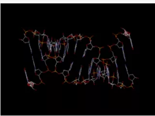
The structure of DNA is illustrated by a right handed double helix, with about 10 nucleotide pairs per helical turn. Each spiral strand, composed of a sugar phosphate backbone and attached bases, is connected to a complementary strand by hydrogen bonding (non- covalent) between paired bases, adenine (A) with thymine (T) and guanine (G) with cytosine (C). Adenine and thymine are connected by two hydrogen bonds (non-covalent) while guanine and cytosine are connected by three. • This structure was first described by James Watson and Francis Crick in 1953.
DNA Double Helix • DNA is a normally double stranded macromolecule. Two polynucleotide chains, held together by weak thermodynamic forces, form a DNA molecule. • Structure of DNA Double Helix • Features of the DNA Double Helix • Two DNA strands form a helical spiral, winding around a helix axis in a right-handed spiral • The two polynucleotide chains run in opposite directions • The sugar-phosphate backbones of the two DNA strands wind around the helix axis like the railing of a sprial staircase • The bases of the individual nucleotides are on the inside of the helix, stacked on top of each other like the steps of a spiral staircase. • As the phosphodiester bonds link carbon atoms number 3′ and number 5′ of successive sugar residues, one end of each DNA strand, the so-called 5′ end, will have a terminal sugar residue in which carbon atom number 5′ is not linked to a neighboring sugar residue . The other end is defined as the 3′ end because of a similar absence of phosphodiester bonding at carbon atom number 3′ of the terminal sugar residue. The two strands of a DNA duplex are said to be antiparallel because they always associate (anneal) in such a way that the 5′ → 3′ direction of one DNA strand is the opposite to that of its partner
Where? • In eukaryotes : nucleus and in mitochondria http://micro.magnet.fsu.edu
Types/Conformation • DNA can adopt different types of helical structure. • A-DNA and B-DNA are both right-handed helices (ones in which the helix spirals in a clockwise direction as it moves away from the observer). They have respectively 11 and 10 base pairs per turn. • Z-DNA is a left-handed helix which has 12 base pairs per turn. Under physiological conditions, most of the DNA in a bacterial or eukaryotic genome is of the B-DNA form in which each helical strand has a pitch (the distance occupied by a single turn of the helix) of 3.4 nm.
A B Z
DNA Discovery • The German biochemist FrederichMiescher first observed DNA in the late 1800s • autumn of 1951, James Watson and Francis Crick started work on unravelling the structure of DNA. • In April 1953 Watson and Crick published their structure - the now famous double helix • 1962 they received the Nobel prize
Crackers of the DNA code • . James Watson and Francis Crick, Science Museum, UK
Biological Function of DNA • DNA stores an organism's genetic information and controls the production of proteins and is thus responsible for the biochemistry of an organism • The actual genetic coding is due to the sequence of bases on the DNA strand. Every amino acid in every protein is coded by sequences of 3 bases • Each DNA sequence that contains instructions to make a protein is known as a gene. The size of a gene may vary greatly, ranging from about 1,000 bases to 1 million bases in humans. • The complete DNA instruction book, or genome, for a human contains about 3 billion bases and about 20,000 genes on 23 pairs of chromosomes
Initiation • AT rich regions . Why? • Pre-replication Complex: Helicase SSBP (Single Stranded Binding Proteins) Primase DNA Holoenzymes (Polymeraze) • Origion of replication • Replication Fork Replication DNA Polymerase, can only synthesize new DNA from the 5’ to 3’ (of the new DNA). Leading Strand DNA ligase, is used to connect the so-called Okazaki fragments. Lagging StrandTerminationThe sticking out 3’ end consists of noncoding DNA called the telomere, which can be simply cut off.
Enzymes of DNA Replication • Helicase: Unwounds a portion of the DNA Double HelixRNA Primase: Attaches RNA primers to the replicating strands.DNA Polymerase delta : Binds to the 5' - 3' strand in order to bring nucleotides and create the daughter leading strand.DNA Polymerase epsilon: Binds to the 3' - 5' strand in order to create discontinuous segments starting from different RNA primers.Exonuclease (DNA Polymerase I): Finds and removes the RNA Primers DNA Ligase: Adds phosphate in the remaining gaps of the phosphate - sugar backboneNucleases: Remove wrong nucleotides from the daughter strand.
Semiconservative • Semidiscontinuous • Replication bubbles
DNA transcription • http://www.youtube.com/watch?v=teV62zrm2P0 • C:\Documents and Settings\Luqshal\My Documents\My Videos\RealPlayer Downloads\DNA replication - Google Video.flv • C:\Documents and Settings\Luqshal\My Documents\My Videos\RealPlayer Downloads\DNA Replication.flv
Genetic information is encoded by the linear sequence of bases in the DNA strands (the primary structure). Consequently, two DNA strands of a DNA duplex are said to have complementary sequences (or to exhibit base complementarity) and the sequence of bases of one DNA strand can readily be inferred if the DNA sequence of its complementary strand is already known. It is usual, therefore, to describe a DNA sequence by writing the sequence of bases of one strand only, and in the 5′ → 3′ direction. This is the direction of synthesis of new DNA molecules during DNA replication, and also of the nascent RNA strand produced during transcription . However, when describing the sequence of a DNA region encompassing two neighboring bases (really a dinucleotide) on one DNA strand, it is usual to insert a ‘p' to denote a connecting phosphodiester bond [e.g. CpG means that a cytidine is covalently linked to a neighboring guanosineon the same DNA strand, while a CG base pair means a cytosine on one DNA strand is hydrogen-bonded to a guanine on the complementary strand .