Download
1 / 108
1.1k likes | 1.36k Views
Acute Coronary syndromes. Yael Moussadji Aug 21, 2008. Objectives. Diagnosis of ACS in the ED Risk Stratification Cardiac markers ECG Risk Scores Management UA/NSTEMI STEMI Complications. Pathophysiology. Definitions. Case 1.

E N D
Acute Coronary syndromes Yael Moussadji Aug 21, 2008
Objectives • Diagnosis of ACS in the ED • Risk Stratification • Cardiac markers • ECG • Risk Scores • Management • UA/NSTEMI • STEMI • Complications
Case 1 • 54 y/o male with 2 hours of exertional retrosternal burning CP • No previous episodes of pain • Feels slightly SOB • VSS, exam normal • ECG non-specific, TnT neg • You ask CCU to see because you are concerned re the possibility of an ACS (UA) • The CCU res asks, does he have any risk factors?
Question 1 Are cardiac risk factors useful in evaluating the risk of ACS?
Post-hoc analysis of 10,806 ED visits for ACS using the i*trACS registry for ED visits for ACS • ACS defined as need for 30-day revascularization (PTCA or CABG), or death or AMI with positive cardiac enzymes at hospitalization • Cardiac RF were diabetes, HTN, dyslipidemia, smoking, + family history of CAD; cardiac RF burden defined as number of RF present • Analysis stratified by age; <40, 40-65, >65
Conclusions • In patients over 40, cardiac risk factor burden is of limited clinical value in the diagnosis of ACS • In patients under 40, cardiac RF useful if there are none (-LR 0.17) or if there are 4 or more (+LR 7.39)
Case 2 • 61 y/o female with 45 minutes of sharp left sided pleuritic chest pain • Feels nauseated, slightly diaphoretic • Pain is radiating to her left shoulder • No PMHx, no DVT/PE risk factors • Cardiac Risk factors: Who cares? • Vital signs are normal, ECG nonspecific, enzymes pending
Question 2 How useful are clinical features in the diagnosis of acute, undifferentiated chest pain?
Measured the predictive value and diagnostic performance of clinical features used to diagnose ACS in undifferentiated CP • Clinical features were prospectively recorded on a standard form for 893 patients presenting to the ED; 3.8% had an MI and 9.1% had ACS • Six month follow-up for adverse events • Tested the power of each feature to predict AMI (WHO criteria) and ACS (cardiac testing, AMI, death, or revascularization within 6 months
Conclusions • Features useful in the diagnosis of AMI were exertional pain (LR 2.35), pain radiating to the shoulder or both arms (LR 4.07), and chest wall tenderness (LR 0.3) • Features useful in the diagnosis of ACS were exertional pain (LR 2.06), pain radiating to the shoulder, left arm, or both arms (LR 1.62) • Location, quality, and presence of N/V or diaphoresis were not predictive
Case 3 • A 57 y/o male with no PMHx presents to the ED with CP • Pain has been intermittent for 2 weeks, and is described as pleuritic and exertional; occational nausea is noted • Physical exam is unremarkable • Patient’s pain resolved spontaneously prior to medical therapy, and he is pain free when you see him
Case 3 continued • Enzymes were negative • Patient was discharged home with instructions to return if worse, and referral to C-era. • 24 hours later, the patient returns to emerg with ongoing chest discomfort, nausea, and diaphoresis
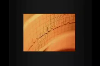
Question 3 What is the predictive and prognostic value of the ECG in patients with ACS?
Definitions • Non-specific ST and T wave changes • ST segment depression or elevation of < 1mm with or without an abnormal T wave • T wave may have altered morphology and/or blunted, flattened, or biphasic configuration without inversion or hyperacuity • Normal • Absence of NSSTTW, AV block, intraventricular conduction delay, repolarization changes, and rhythms other than NSR
ECG Findings in ACS • In a study of adult CP patients in the ED, 1% of patients with anormal ECG had a final diagnosis of AMI, and 4% had a final diagnosis of UA • In another study, of patients with classic angina on history and a normal ECG, 3% had a final diagnosis of AMI • 3-4% of patients with AMI and over 20% of patients with an ACS (NSTEMI/UA) have NSSTTW findings • Therefore, of all patients with ACS, one fifth will show a normal or non-specific ECG in the ED
Of 202 chest pain patients presenting to the ED with STE, 15% had an AMI • LVH was the most common cause of STE (25%), followed by LBBB (15%) and AMI (15%) • 12% had BER, 5% had RBBB, and 5% had nonspecific BBB • Other less common diagnoses were LVA, pericarditis, and paced rhythm
Prognostic Value of Admission ECG in ACS • A retrospective analysis of GUSTO-IIb trial • Over 12,000 patients who had ACS confirmed on ECG • 22% had T wave inversion, 28% had STE, 35% had STD, and 15% had a combination of the above • 30 day incidence of death or MI was 5.5% in those with T wave inversions, 9.4% in those with STE, 10.5% in those with STD, and 12.4% in those with a STE + STD • In another study of 205 consecutive patients with UA/NSTEMI, STE of > 0.5mm in aVR was found to be a strong predictor of 30-day mortality, even in patients with low TIMI risk scores
GUSTO 2B: ST DepressionA High Risk Finding ST P 0.001 ST T-wave inversion CM Gibson 2002
ECG Pearls • 50% of patients with AMI will have a clearly diagnostic ECG at presentation (STE or STD) • ST segment elevation identifies those who benefit from reperfusion therapy (lytics) • Mortality increases with the number of leads showing STE • Other important predictors of mortality include LBBB and anterior location • Reciprocal changes are seen in 70% of inferior and 30% of anterior MIs, which demonstrates over 90% specificity and PPV for AMI • RV infarcts complicate 40% of inferior AMIs
Question 4 So, if risk factors, clinical features, and ECG’s are not always helpful, how many patient’s with ACS are missed, and what are their characteristics?
Analyzed clinical data from a multicentre prospective trial of over 10,000 patients with chest pain suggestive of ACS • 17% ultimately met the criteria for ACS (8% had AMI and 9% had UA) • 2.1% of those with AMI and 2.3% of those with UA were mistakenly discharged from the ED
Missed diagnosis of ACS • Acute ischemia • Women <55 • Non-white • SOB as chief symptom • Normal or non-diagnostic ECG • AMI • Non-white • Normal or non-diagnostic ECG
Conclusion • Percentage of patients who get discharged home is low, but discharge of these patients may be associated with increased mortality • Failure to make a diagnosis is related to race, gender, and lack of typical features on ECG
Case 4 • 83 y/o male with known renal insufficiency, baseline Cr 150 • Presents with vague intermittent CP of 2 days duration, no associated symptoms • PMhx significant for HTN, previous MI and PCTA 10 years ago • ECG non-diagnostic (no acute changes from baseline) • TnT 0.11 • CCU res says “it’s elevated because of his renal failure”
Question 5 Can you diagnose ACS based on an elevated TnT in a patient with renal failure?
Analyzed outcomes in over 7000 patients enrolled in the GUSTO IV trial • Assessed baseline TnT level (considered abnormal if >0.1 ng/mL) and Cr clearance • Primary end point was death or MI at 30 days • An elevated TnT level was predictive of death of MI, even among patients with a Cr clearance in the lowest quartile • Cardiac troponin is predictive of short term prognosis in patients with ACS regardless of their level of Cr clearance
Cardiac Troponin • Due to near absolute specificity for myocardial tissue and high sensitivity for microscopic zones of myocardial necrosis, cardiac troponins are the preferred biomarker for diagnosing MI • Onset 3-6 hours • Peak 12-18 hours • Elevated for 5-7 days
Examined the TnT, CK-MB, and ECG abnormalities for risk stratification in patients with ACS within 12 hours on onset of symptoms • Use logistic regression to predict outcome • Mortality was significantly higher in the group with Tn >0.1 ng/mL (ARR 8%) • TnT was the variable most strongly related to 30 day mortality, followed by ECG category and the CK-MB level • TnT is a powerful independent predictor of mortality in patients who present with ACS
Prospectively examined 733 patients with acute CP < 12 hours without STE; Tn was measured at least twice on arrival and 4-6 hours later so that one sample was taken at least 6 hours after the onset of pain • TnT was positive in 16% of patients, and 94% of patients who eventually evolved into an AMI • Among patients with UA, TnT was positive in 20% • TnT was a strong independent predictor of cardiac events • The event rate for patients with negative Tn T was 1.1%
Risk Stratification • 2 questions • What is the likelihood that the presenting symptoms represent ACS? • What is the likelihood of adverse outcome • Risk stratification process is challenging given then presence of risk factors is an unreliable determinant of ACS, and the ECG and Tn are not very sensitive for UA • 2007 ACC/AHA Update to the guidelings for UA/NSTEMI are helpful
Use of Risk Stratification Tools • 2002 Guidelines state that tools such as the TIMI Risk Score can be helpful adjuncts • Since 2002, data from a unselected ED chest pain population have validated its utility • Other recommended tools include the GRACE (Global Registry of Acute Coronary Events) Risk Score and the PURSUIT (Platelet Glycoprotein IIb/IIIa in Unstable Angina: Receptor Suppression Using Integrilin Therapy) risk model • A study comparing the 3 showed good predictive accuracy for death at 1 year and MI • However, these tools were developed using population based models and may not be reliable for individual patients; they do not replace clinical judgement
Two phase 3 international, randomized, double-blinded trials (TIMI 11B, ESSENCE) • A total of 1957 with UA/NSTMEI who were assigned to receive UFH in TIMI 11B(test cohort) • 3 validation cohorts were the UFH group in ESSENCE and both enoxaparin groups (total of over 5000 patients) • Risk score was derived from test cohort using multivariate logistic regression, assinging a value of 1 when risk factor present, and 0 when absent • Outcomes were at least 1 component of the primary end point (mortality, MI, urgent revascularization)
Results • TIMI Risk Score • Age > or = 65 • 3 or more risk factors for CAD • Prior stenosis of 50% or more • ST segment deviation at presentation • At least 2 anginal events in 24 hours • Use of ASA in prior 7 days • Elevated serum cardiac markers