Download
1 / 59
600 likes | 882 Views
Preeclampsia in the Parturient Implications in Anesthesia. Robyn C. Ward, CRNA, MS LCDR, NC, USN Naval Medical Center San Diego. www.anaesthesia.co.in anaesthesia.co.in@gmail.com. Objectives . Discuss preeclampsia: risk factors, pathophysiology, obstetric/anesthetic management

E N D
Preeclampsia in the ParturientImplications in Anesthesia Robyn C. Ward, CRNA, MS LCDR, NC, USN Naval Medical Center San Diego www.anaesthesia.co.inanaesthesia.co.in@gmail.com
Objectives • Discuss preeclampsia: risk factors, pathophysiology, obstetric/anesthetic management • Discuss eclampsia • Discuss HELLP syndrome
Introduction • Preeclampsia is a major cause of maternal and perinatal morbidity and mortality • Direct fetal effects • Indirect effects due to preterm deliveries • Affects ~ 5% of all pregnancies • 35-300 deaths per 1000 births (twice that of normotensive pregnancies)
Classification – 4 Categories • Gestational hypertension • Chronic hypertension • Chronic hypertension w/ superimposed preeclampsia • Preeclampsia – mild, moderate, or severe
Gestational Hypertension • Transient HTN of BP > 140/90 without proteinuria or end-organ damage • May occur late in pregnancy, during labor, or within 24 hrs postpartum • BP returns to normal within 10 days postpartum
Chronic Hypertension • Begins prior to pregnancy • BP > 140/90 • Occurs prior to 20th week gestation • Not associated with proteinuria or end-organ damage • Continues well after delivery
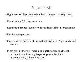
Preeclampsia – Mild to Severe • Occurs after 20 weeks gestation • Triad: • Hypertension • Proteinuria • Edema
Preeclampsia - Mild • Hypertension: SBP > 140 mm Hg, DBP > 90 mm Hg • Proteinuria: > 300 mg/24 hr • Generalized edema
Preeclampsia - Severe • Hypertension: SBP > 160 mm Hg, DBP > 110 mm Hg • Proteinuria: > 5 g in 24 hr • Generalized edema • Evidence of end-organ damage: • Oliguria: U/O < 400 ml/24 hr and/or elevated serum creatinine • Cerebral edema w/ visual disturbances (blurred vision) or headache
Preeclampsia – Severe (cont) • Evidence of end-organ damage (cont.): • Pulmonary edema • Hepatocellular dysfunction • Elevated liver enzymes • Epigastric or RUQ pain • HELLP syndrome • Intrauterine growth restriction or oligohydramnios
Risk Factors • Maternal Pre-existing Conditions • Maternal Specific Factors • Partner-Related Factors • Pregnancy-Associated Factors
Maternal Pre-existing Conditions • Chronic hypertension • Renal disease • Diabetes mellitus • Obesity
Maternal Specific Factors • Advance maternal age > 40 • Familial predisposition • Nulliparity and young age • History of preeclampsia
Partner-Related Factors • Paternal antigens • Immune maladaptation • Trophoblastic invasion at 12 weeks • Second invasion at 14-16 weeks changes spiral arteries from high to low resistance vessels; prostacyclin and nitric oxide are released from endethelium • Second invasion fails in preeclampsia
Pregnancy-Associated Factors • Multiple gestation – increased risk • Congenital anomalies • Antepartum urinary tract infection
Pathophysiology • Immunologic factors • Genetic factors • Endothelial factors • Platelet factors • Calcium
Immunologic Factors • Fifty percent genes from father • Fetal trophoblast invades maternal deciduas after implantation • Second trophoblastic invasion at 14-16 weeks normally disrupts maternal spiral arteries changing them to low resistance vessels • This 2nd wave of invasion fails in preeclampsia
Genetic Factors • Possibly recessive genetic inheritance • Variant angiotensinogen gene T 235 – if homozygous, 20 fold increased risk for preeclampsia
Endothelial Factors • Vascular endothelial damage • Occurs in placental deciduas, renal microvasculature and, potentially, hepatic, myocardial and cerebral circulations • Failure of trophoblast invasion causes increases production of free radicals which exacerbate endothelial damage • Overall result is deficiency of prostacyclin (vasodilator) in relation to thromboxane (vasoconstrictor)
Platelet Factors • Surface mediated platelet activation occurs in absence of prostacyclin • Favors platelet adhesion to damaged surface lining of spiral arteries • Further platelet aggregation promoted by platelet release of dense granules, including TXA2 and 5HT
Platelet Factors (cont.) • Mild preeclampsia, 5HT binds to endothelial 5HT-1 receptors • stimulates partial recovery of endothelial prostacyclin and nitric oxide release • Prostacyclin stimulates renin angiotensin aldosterone system, which improves placental perfusion by inducing maternal HTN
Platelet Factors (cont.) • Severe preeclampsia, serotonin cannot interact with 5HT-1 receptors on endothelium • Binds instead to 5HT-2 receptors on vascular smooth muscle cells and platelet membranes • Results in worsening HTN and intravascular thrombosis
Calcium • Calcium increases slowly throughout normal pregnancy to maintain vascular tone • In preeclampsia, this increase is much greater by 3rd trimester due to increased cellular membrane permeability
Clinical Manifestations • Cardiovascular • Respiratory • Neurologic • Renal • Hematologic • Hepatic • Uteroplacental unit
Cardiovascular • HTN • Intravascular volume depletion • shift of intravascular fluid and protein to extravascular space due to decreased colloid oncotic pressure and increased capillary permeability (due to endothelial cell damage)
Respiratory • Upper AW narrowing from pharyngolaryngeal edema • difficulty in intubation • Pulmonary edema in 3% of cases • Increased capillary permeability • Decreased colloid oncotic pressure • LV dysfunction • Excessive fluid administration
Neurologic • Cerebral irritation due to CNS edema • Headache – 40% of patients • Nausea, excitability, apprehension and visual disturbances (blurred vision) • Hyperreflexia/Clonus – seizures if progresses to eclampsia • Intracranial hemorrhage is leading cause of maternal death from preeclampsia
Renal • Diminished renal perfusion and glomerular filtration • Proteinuria • Decreased uric acid clearance • Oliguria < 400 ml/24 hrs • Elevated serum creatinine
Hematologic • Elevated Hct secondary to plasma volume contraction • Thrombocytopenia in 30% of patients • PLT < 100K correlates with severe disease • Activation of coagulation cascade can occur • Increased incidence of placental abruption which can result in DIC
Hepatic • Impaired liver function can be caused by: • Periportal hepatic necrosis • Subscapular hemorrhages • Fibrin deposition in hepatic sinusoids • Elevated liver enzymes (HELLP)
Uteroplacental Unit • Uteroplacental insufficiency due to increased vascular resistance, decreased plasma volume, and increased blood viscosity • Compromises uterine blood flow by 50-70%
Treatment • Obstetric management • Adequate fetal surveillance • Antihypertensive management • Anticonvulsant therapy • Anesthetic management of labor • Safe analgesia for labor and anesthesia for delivery
Obstetric Management • Expectant management (< 34 weeks) • Bed rest and sedation • Antihypertensive therapy • Monitoring of weight, U/O and DTR’s • MgSO4 for seizure prophylaxis
Obstetric Management • Aggressive management • Induction of labor/delivery within 48-72 hours • Delivery is only curative treatment • If possible, time allowed for 2 doses of steroids to mother to promote fetal lung maturity • Indications for immediate delivery: • Uncontrolled HTN > 160/110 • Oliguria/renal dysfunction • Liver dysfunction • Imminent eclampsia • Pulmonary edema • Fetal compromise
Fetal Surveillance • Fetal ultrasound • Continuous electronic fetal monitoring • Monitor for: • Loss of beat-to-beat variability of FHR and periodic late decelerations
Antihypertensive Management • Goal: control HTN and maintain uteroplacental perfusion • Acute therapy: Long Term: • Hydralazine Alpha methyl dopa • Labetalol (IV) Labetalol (po) • Nifedipine • Nitroglycerin • Nitroprusside
Anticonvulsant Therapy • MgSO4 = first line agent in U.S. • Cerebral vasodilator • Central anticonvulsant effect • Peripheral vasodilator effect • Other: • Phenytoin (Dilantin) • Diazepam
Magnesium • Dose • 4-6 g over 20 min w/ infusion of 1-2 g/hr • Therapeutic range = 4-8 mg/dl • Side effects: sedation, skeletal/muscle weakness, decreased uterine activity, decreased FHR variability, prolonged labor, uterine atony, prolongation of NM blockade, neonatal hypotonia
Magnesium (cont.) • Toxicity: • 4-8 mg/dl = therapeutic • 10-12 mg/dl = loss of DTRs • 12-15 mg/dl = respiratory arrest • 20-25 mg/dl = cardiac arrest • Treatment: • Calcium gluconate 10 ml of 10% solution IV over 2 minutes • Oxygen • Mechanical ventilation if necessary
Anesthetic Management of Labor • Preanesthetic assessment: • Airway • Aspiration prophylaxis • Auscultation of lungs • Fluid balance • Hemodynamic status • Left uterine displacement • Renal function • Coagulation status
Analgesia for Labor • Continuous lumbar epidural – Advantages: • Decreased circulating catecholamines • Decreased uterine vascular resistance • Improved uteroplacental blood flow • Avoids risk of general anesthesia
Epidural Placement • R/O coagulopathy, LUD, oxygen, continuous fetal monitoring • Careful crystalloid preload (250-500 ml) • Local anesthetic: Bupivicaine (slow onset) • Epinephrine: consider avoiding • Slow, incremental dosing • Ephedrine (in smaller doses) for hypotension < 20% of baseline
Anesthesia for Delivery • Non-emergent C-section: • Epidural anesthesia: thought to allow for incremental dosing, potentially avoiding precipitous hypotension • Spinal anesthesia: recent retrospective study (Hood & Curry, 1999) found no difference in hemodynamic changes after spinal or epidural anesthesia • Conclusion: spinal is safe alternative to epidural w/ added advantage of quicker onset and better quality of sensory blockade especially in urgent situations
Anesthesia for Delivery • Emergent C-section • Epidural – previously placed, well functioning • Spinal – if no epidural placed and if FHR stable • General anesthesia: • Coagulopathy • Patient refusal of regional • Fetal bradycardia prohibits placement in time
Eclampsia • Convulsions that occur during pregnancy in a woman whose condition also meets criteria for preeclampsia and who has no other previous neurologic disease or diagnosis of epilepsy • Etiology uncertain: cerebral edema, ischemia • Tonic-clonic in nature • Life threatening emergency
Eclampsia (cont.) • 50% have evidence of severe preeclampsia • Classical presentation of preeclampsia may be absent or mildly abnormal in 30% of patients
Management of Eclampsia • Goals: • Stabilization of the mother/seizure control • MgSO4 therapy: 4-6 g over 20 min followed by infusion of 1-3 g/hr, OR • Thiopental or diazepam followed by MgSO4 infusion • Avoid anticonvulsant polypharmacy • Airway management • Avoiding aspiration
Obstetric Management of Eclampsia • Factors to consider: patient’s condition, gestational age, cervical exam, and fetal well being • Induction of labor if patient is seizure free, stable, favorable cervix, and reassuring fetal heart tracing • Urgent c-section if patient continues to have breakthrough seizures
Anesthetic Management of Eclampsia • Aspiration precautions • Careful airway assessment • Guidelines for use of regional vs. general anesthesia similar to patients with severe preeclampsia
ABC’s of Seizure Control • Airway • Breathing • Circulation • Drugs • Blood Pressure