Download
1 / 59
590 likes | 810 Views
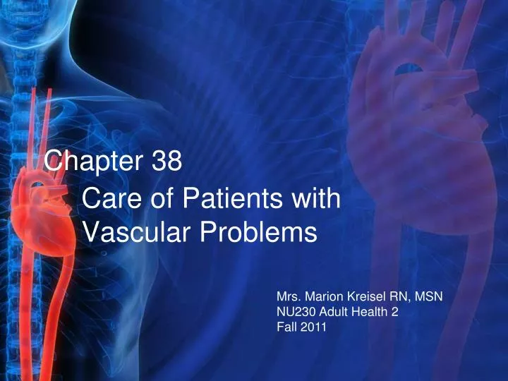
Chapter 38. Care of Patients with Vascular Problems. Mrs. Marion Kreisel RN, MSN NU230 Adult Health 2 Fall 2011. Arteriosclerosis and Atherosclerosis. Arteriosclerosis—thickening or hardening of the arterial wall often associated with aging.

E N D
Chapter 38 Care of Patients with Vascular Problems Mrs. Marion Kreisel RN, MSN NU230 Adult Health 2 Fall 2011
Arteriosclerosis and Atherosclerosis • Arteriosclerosis—thickening or hardening of the arterial wall often associated with aging. • Atherosclerosis—type of arteriosclerosis involving the formation of plaque within the arterial wall. • Etiology and genetic predisposition: • Factors related to atherosclerosis include obesity, lack of exercise, smoking, and stress.
Laboratory Assessment • Lipid level, including cholesterol and triglycerides, elevated • HDL and LDL • High serum levels of homocysteine can allow cell walls to become vulnerable to plaque buildup
Interventions • Evaluation of total serum cholesterol levels and lifestyle changes • Nutrition therapy • Smoking cessation • Exercise • National Cholesterol Education Program (NCEP) • Therapeutic Lifestyle Change (TLC) diet
Drug Therapy • HMG-CoA reductase inhibitors (statins) • Fibrinic acids • Zetia • Omacar
Hypertension • Hypertension—systolic blood pressure ≥ 145 mm Hg and/or diastolic blood pressure ≥ 90 mm Hg in people who do not have diabetes mellitus. • Patients with DM should have a BP below 130/90. • “Normal” adult systolic BP less than 120; diastolic less than 80.
Hypertension (Cont’d) • Prehypertensive systolic 120 to 139 and diastolic 80 to 89. • Isolated systolic hypertension. • Malignant hypertension is a severe type of elevated BP that rapidly progresses.
Essential Hypertension • Age greater than 60 years • Family history of hypertension • Excessive calorie consumption • Physical inactivity • Excessive alcohol intake • Hyperlipidemia • African-American ethnicity • High intake of salt or caffeine
Essential Hypertension (Cont’d) • Reduced intake of K, Ca, or Mg • Obesity • Smoking • Stress • Monitor for increase in BUN (10-20mg.d/L) and Serum Creatinine (0.5-1.2mg/dL) Levels
Secondary Hypertension • Renal disease • Primary aldosteronism • Pheochromocytoma • Cushing’s syndrome • Medications
Assessment • Patient history • Physical assessment • Psychological assessment • Diagnostic assessment
Knowledge Deficit • Interventions include: • Sodium restriction • Weight reduction • Moderation of alcohol intake • Exercise • Relaxation techniques • Tobacco and caffeine avoidance
Drug Therapy • Diuretics: 3 basic types used to decrease b/p • Thiazides: hydrochlorothiazide (HydroDIURIL, Urozide. Promote NA+ & K+ excreation • Loop: Furosemide (Lasix) promote Na+ & K+ excreation • K+ Sparing: spironolactone(Aldactone, Novospiroton) inhibits Na+ reabsorption and retains K+ • Calcium channel blockers: Verapamil hydrochloride (Calan) & amlodipine (Norvasc) Vasodilation • ACE inhibitors: captopril (Capoten) & enalapril (Vasotec). decrease vasoconstriction and control B/P (cough) • VERY IMPORTANT EDUCATION ABOUT MEDS! • WATCH FOR OTROSTATIC HYPOTENSION!
Drug Therapy Continued • Angiotensin II receptor antagonists: • Aldosterone receptor antagonists • Beta-adrenergic blockers: drug of choice for patients with ischemic heart disease. • Renin inhibitors: new category of drugs mild to moderate HTN enzyme produced by kidneys to cause vasoconstriction therefore they inhibit that mechanism • Central alpha agonists • Alpha-adrenergic agonists
Risk for Ineffective Therapeutic Regimen Management • Interventions include: • Teach medication compliance, usually for the rest of life. • Discuss goals of therapy, potential side effects, and how to identify potential problems. • Assist patient to understand therapeutic regimen. • Discuss consequence of noncompliance.
Peripheral Arterial Disease • Disorders that alter the natural flow of blood through the arteries and veins of the peripheral circulation. Extreme lose of feeling can occur so pt education very important • Can lead to a DVT. Pt will be on anticoagulation therapy and will need lots of pt education.
Physical Assessment • Intermittent claudication • Pain that occurs even while at rest; numbness and burning • Inflow disease discomfort in the lower back, buttocks, or thighs • Outflow disease burning or cramping in the calves, ankles, feet, and toes
Physical Assessment(Cont’d) • Hair loss and dry, scaly, pale or mottled skin and thickened toenails • Severe arterial disease—extremity is cold and gray-blue or darkened; pallor may occur with extremity elevation; dependent rubor; and/or muscle atrophy
Diagnostic Assessments • Imaging assessment • Other diagnostic tests: • Ankle-brachial index (ABI) • Exercise tolerance testing • Plethysmography
Nonsurgical Management • Exercise • Positioning • Promoting vasodilation • Drug therapy • Percutaneous transluminal angioplasty • Laser-assisted angioplasty • Atherectomy
Surgical Management • Aortoiliac and aortofemoral bypass surgery
Surgical Management • Preoperative • Intraoperative
Surgical Management (Cont’d) • Postoperative care: • Assessment for graft occlusion • Promotion of graft patency • Treatment of graft occlusion • Monitoring for compartment syndrome • Assessment for infection
Acute Peripheral Arterial Occlusion • Embolus—the most common cause of occlusions, although local thrombus may be the cause • Assessment—pain, pallor, pulselessness, paresthesia, paralysis, poikilothermia • Drug therapy • Surgical therapy • Nursing care
Aneurysms of Central Arteries • Aneurysm—a permanent localized dilation of an artery, enlarging the artery to twice its normal diameter • Fusiform aneurysm • Saccular aneurysm • Dissecting aneurysm (aortic dissection) • Abdominal aortic aneurysm • Thoracic aortic aneurysm
Assessment of Abdominal Aortic Aneurysm (AAA) • Pain related to AAA is usually steady with a gnawing quality, is unaffected by movement, and may last for hours or days. • Pain is in the abdomen, flank, or back. • Abdominal mass is pulsatile. • Rupture is the most frequent complication and is life threatening.
Assessment of Thoracic Aortic Aneurysm • Assess for back pain and manifestation of compression of the aneurysm on adjacent structures. • Assess for shortness of breath, hoarseness, and difficulty swallowing. • Occasionally a mass may be visible above the suprasternal notch. • Sudden excruciating back or chest pain is symptomatic of thoracic rupture. • PLAN FOR IMMEDIATE SURGERY TO SAVE PT LIFE!
Diagnostic Assessment • X-ray “eggshell” appearance • CT • Aortic arteriography • Ultrasonography
Nonsurgical Management • Monitor the growth of the aneurysm. • Maintain BP at a normal level to decrease the risk of rupture.
Abdominal Aortic Aneurysm Resection • Preoperative care • Operative procedure • Postoperative care: • Monitor vital signs • Assess for complications such as decrease u/o. If this happens MD will order kidney function tests • Assess for signs of graft occlusion or rupture
Thoracic Aortic Aneurysm Repair • Preoperative care • Operative procedure • Postoperative care assessments: • Vital signs • Complications • Sensation and motion in extremities • Respiratory distress • Cardiac dysrhythmias
Endovascular Repair of Abdominal Aortic Aneurysm • Patients selected for endovascular repair are generally at high risk for major abdominal surgery • Various designs • Benefits of endovascular repair • Complications of endovascular repair
Aneurysms of the Peripheral Arteries • Femoral and popliteal aneurysms • Symptoms—limb ischemia, diminished or absent pulses, cool to cold skin, and pain • Treatment—surgery • Postoperative care—monitor for pain
Aortic Dissection • May be caused by a sudden tear in the aortic intima, opening the way for blood to enter the aortic wall • Pain described as tearing, ripping, and stabbing
Aortic Dissection (Cont’d) • Emergency care goals include: • Elimination of pain • Reduction of blood pressure • Decrease in the velocity of left ventricular ejection • Nonsurgical treatment • Surgical treatment
Buerger’s Disease • Thromboangiitis obliterans—relatively uncommon occlusive disease limited to the medium and small arteries and veins • Often identified with tobacco smoking • Nursing interventions
Other Disorders • Subclavian steal occurring from artery occlusion or stenosis • Thoracic outlet syndrome resulting in arterial wall damage • Popliteal entrapment
Raynaud’s Phenomenon • Caused by vasospasm of the arterioles and arteries of the upper and lower extremities • Drug therapy—Procardia, Cyclospasmol, and Dibenzyline • Lumbar sympathectomy • Reinforcement of patient education; restriction of cold exposure
Venous Thromboembolism • Thrombus—a blood clot • Thrombophlebitis • Deep vein thrombosis (DVT) • Pulmonary embolism • Virchow’s triad • Phlebitis
Assessment • Calf or groin tenderness or pain • Sudden onset of unilateral swelling of the leg • Checking Homans’ sign—not advised • Localized edema • Venous flow studies—venous duplex ultrasonography • MRI • D-dimer
Nonsurgical Management • Rest, drug therapy, preventive measures • Drug therapy includes: • Unfractionated heparin therapy • Low–molecular weight heparin • Warfarin therapy • Thrombolytic therapy
Surgical Management • Thrombectomy • Inferior vena caval interruption • Ligation or external clips
Venous Insufficiency • Result of prolonged venous hypertension, stretching veins and damaging valves • Stasis dermatitis, stasis ulcers • Management of edema • Management of venous stasis ulcers • Drug therapy • Surgical management