Download
1 / 196
1.98k likes | 2.25k Views
Chapter 1 Protein. Contents. Chemical components Molecular structures Biological functions Structure-function relationship Physical and chemical properties Exploration of proteins Proteomics: a new frontier. What are p roteins?.

E N D
Chapter 1 Protein
Contents • Chemical components • Molecular structures • Biological functions • Structure-function relationship • Physical and chemical properties • Exploration of proteins • Proteomics: a new frontier
What are proteins? Proteins are macromolecules composed of amino acids linked together through peptide bonds.
How are about proteins? • the most widely distributed biomolecules • the most abundant biomolecules (45% of human body) • the most complex biomolecules • the most diversified biological functions
Section 1 Chemical Components of Proteins
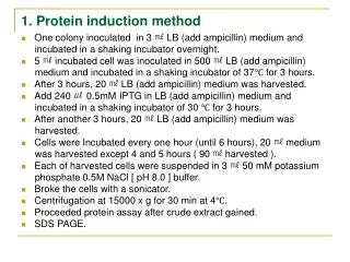
Components of proteins • major elements C (50~55%), H (~7%), O (19~20%), N (13~19%),S (~4%) • trace elements P, Fe, Cu, Zn, I, …
The average nitrogen content in proteins is about 16%, and proteins are the major source of N in biological systems. • The protein quantity can be estimated. protein in 100g sample = N per gram x 6.25 x 100
§1.1 Amino Acids • The basic building blocks of proteins • About 300 types of AAs in nature, but only 20 types are used for protein synthesis in biological systems. • A amino group, a carboxyl group, a H atom and a R group are connected to a C atom. • The C atom is an optically active center.
Molecular weight Dalton: A unit of mass nearly equal to that of a hydrogen atom Gly C2NO2H5 75 Ala C3NO2H7 89 Val C5NO2H11 117 Leu C6NO2H13 131 Ile C6NO2H13 131
§1.1.a Classification • The R groups, also called side chains, make each AA unique and distinctive. • R groups are different in their size,charge,hydrogen bonding capability and chemical reactivity. • Aas are grouped as (1) non-polar, hydrophobic; (2) polar, neutral; (3) basic; and (4) acidic.
Non-polar and hydrophobic AAs • R groups are non-polar, hydrophobic aliphatic or aromatic groups. • R groups are uncharged. • AAs are insoluble in H2O.
Polar and uncharged AAs • R groups are polar: -OH, -SH, and -NH2. • R groups are highly reactive. • AAs are soluble in H2O, that is, hydrophilic.
Basic AAs • R groups have one -NH2. • R groups are positively charged at neutral pH (=7.0). • AAs are highly hydrophilic.
Acidic AAs • R groups have –COOH. • R groups are negatively charged at physiological pH (=7.4). • AAs are soluble in H2O.
Aspartic acid glutamic acid (Asp or D) (Glu or E)
Nomenclature Starting from the carboxyl group, and naming the rest carbon atoms sequentially in Greek letters. -amino-propionic acid -amino--guanidinovaleric acid (alanine) (arginine)
Special amino acids - Gly • optically inactive
Special amino acids - Pro • Having a ring structure and imino group
Special amino acids - Cys active thiol groups to form disulfide bond
§1.2 Peptide §1.2.a Peptide and peptide bond A peptide bond is a covalent bond formed between the carboxyl group of one AA and the amino group of its next AA with the elimination of one H2O molecule.
Peptides can be extended by adding multiple AAs through multiple peptide bonds in a sequential order. dipeptide, tripeptide, oligopeptide, polypeptide AAs in peptides are called as residues.
§1.2.b Biologically active peptides Glutathione (GSH) Glutamic acid cystein glycine
H2O2 2GSH 2H2O GSSG NADP+ GSH peroxidase GSH reductase NADPH+H+ As a reductant to protect nucleic acids and proteins from toxin by discharging free radical or H2O2
Peptides • Peptide hormones secreted from peptidergic neurons or • Somatostatin, Noacosapeptide, Octapeptide, • Thyrotropin-release hormone, Antidiuretic hormone • Neuropeptides responsible for signal transduction • Enkephalin, Endorphin, Dynorphin, Substance P, Neuropeptide Y
thyrotropin-release hormone Pyroglutamic acid histidine prolinamide
Section 2 Molecular Structures of Proteins
Overview • Proteins are composed of AAs. • Distinctive properties of proteins are determined by AA compositions, AA sequences as well as the relative positions of AAs in space. • Proteins need well defined structures to function properly. Their structures are organized in a hierarchy format, that is, primary, secondary, tertiary and quaternary structure.
§2.1 Primary Structure The primary structure of proteins is defined as a linear connection of AAs along the protein chain. It is also called amino acid sequence. The AA sequence must be written from the N-terminus to the C-terminus. Peptidebonds are responsible for maintaining the primary structure.
Primary structure of insulin • Two peptides of 21 and 30 AAs • Two inter-chain -S-S- bonds • One intra-chain -S-S- bond
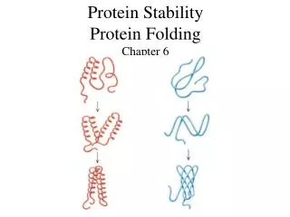
§2.2 Secondary Structure The secondary structure of a protein is defined as a local spatial structure of a certain peptide segment, that is, the relative positions of backbone atoms of this peptide segment.
Repeating units of N(-H), C, and C(=O) constitute the backbone. • H-bonds are responsible for stabilizing the secondary structure. • The side chains are not considered. • -helix -pleated sheet -turn (-bend) random coil
Peptide unit • Six atoms, C-C(=O)-N(-H)-C, constitute a planer peptide unit. • The peptide unit is rigid due to the partial double bond property. • C=O and N-H groups are in trans conformation and cannot rotate around the peptide bond.
Linus Carl Pauling b. 1901, d. 1994 California Institute of Technology, CA The Nobel Prize in Chemistry (1954), “for his research into the nature of the chemical bond and its application to the elucidation of the structure of complex substances” The Nobel Peace Prize (1962)
§2.2.a -helix • A helical conformation is right-handed. • 3.6 AAs per turn and a 0.15 nm vertical distance, creating a pitch of 0.54 nm. • Side chains of AA residues protrude outward from the helical backbone. • The hydrogen-bonds are parallelto the helical axis.
The -CO group of residue n is H-bonded to the -NH group of residue (n+4).
§2.2.b -pleated sheet • An extended zigzag conformation of protein backbones • Protein backbones are arranged side-by-side through H-bonds. • H-bonds are perpendicular to the backbone direction. • The side chains of adjacent AAs protrude in opposite directions. • The adjacent protein backbones can be either parallel or anti-parallel.