Download
1 / 53
580 likes | 938 Views
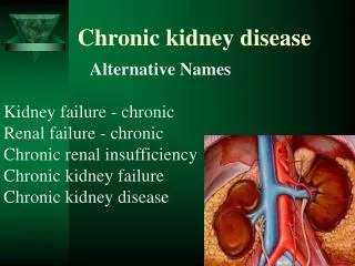
Chronic Kidney Disease. Progressive, irreversible damage to the nephrons and glomeruli. Chronic Kidney Disease. Major causes are. Diabetes and high blood pressure Type 1 and type 2 diabetes mellitus High blood pressure (hypertension) Glomerulonephritis Polycystic kidney disease

E N D
Progressive, irreversible damage to the nephrons and glomeruli Chronic Kidney Disease
Major causes are • Diabetes and high blood pressure • Type 1 and type 2 diabetes mellitus • High blood pressure (hypertension) • Glomerulonephritis • Polycystic kidney disease • Use of analgesics - acetaminophen(Tylenol) and ibuprofen (Motrin, Advil • Clogging and hardening of the arteries(atherosclerosis) • Obstruction of the flow of urine by stones, an enlarged prostate, strictures (narrowings), or cancers. • HIV infection, sickle cell disease, heroin abuse, amyloidosis, kidney stones, chronic kidney infections, and certain cancers.
Kidney functions - monitored regularly • Diabetes mellitus type 1 or 2 • High blood pressure • High cholesterol • Heart disease • Liver disease • Amyloidosis • Sickle cell disease • Systemic Lupus erythematosus • Vascular diseases such as arteritis, vasculitis, or fibromuscular dysplasia • Vesicoureteral reflux (a urinary tract problem in which urine travels the wrong way back toward the kidney) • Require regular use of anti-inflammatory medications • A family history of kidney disease
Protein and waste metabolism accumulates in the blood (azotemia) 90% of kidney function is lost (kidney cannot adequately function) Hypothesis: Nephrons remains intact, others progressively destroyed. Adaptive response maintains function until ¾ are destroyed Hypertrophy continues kidneys begin to lose their ability to concentrate the urine adequately Chronic Renal FailureEnd Stage Renal Disease (ESRD)
Table 1. Stages of Chronic Kidney Disease *GFR is glomerular filtration rate, a measure of the kidney's function.
Modifiable Factors -Diabetic Mellitus -Hypertension -Increase Protein and Cholesterol Intake -Smoking -Use of analgesics Non-Modifiable Factors -Hereditary -Age greater than 60 years old -Gender -Race Decreased renal blood flow Primary kidney disease Damage from other diseases Urine outflow obstruction Chronic Kidney Disease - Pathophysiology BUN Serum Creatinine Decreased glomerular filtration Hypertrophy of remaining nephrons Dilute Polyuria Loss of Sodium in Urine Hyponatremia Inability to concentrate urine Dehydration Further loss of nephron function Loss of nonexcretory renal function 2a Failure to convert inactive forms of calcium Failure to produce eryhtropoietin Impaired insulin action Production of lipids Immune disturbances Disturbances in reproduction Erratic blood glucose levels Advanced atherosclerosis Anemia Pallor Calcium absorption Delayed wound healing Infection Libido Infertility 1
2a 1 Hypocalcemia Osteodystrophy Loss of excretory renal function Excretion of nitrogenous waste Decreased sodium reabsorption in tubule Decreased potassium excretion Decreased phosphate excretion Decreased hydrogen excretion Uremia Hyperkalemia Hyperphosphatemia Metabolic acidosis Water Retention BUN, Creatinine Uric Acid Decreased calcium absorption Hypertension Heart Failure Edema Proteniuria Hypocalcemia Peripheral nerve changes Hyperparathyroidism Decreased potassium excretion Pericarditis Increased potassium CNS changes Pruritus Altered Taste Bleeding Tendencies
Weakness and tiredness/ fatigue. • Nocturia is often an early symptom • Itchiness of the skin which can progressively worsen • Pale skin which is easily bruised • Muscular twitches, cramps and pain • Pins and needles in the hands and feet • Nausea
As the condition worsens the symptoms progress to: • Oedema (swelling of the face, limbs and abdomen) • Oliguria (greatly reduced volume of urine) • Dyspnoea (breathlessness) • Vomiting • Confusion • Seizures • Severe lethargy • Very itchy skin • Breath that smells of ammonia
Associated complications of chronic Kidney Disease would be: • Anaemia, mostly due to deficiency of erythropoietin • Bleeding which is caused by impairment of platelet function • Metabolic Bone Disease (known as Renal Osteodystrophy)
Associated complications of chronic Kidney Disease would be: • Cardiovascular Disease - hypertension, (which may further exacerbate the renal failure) -accelerated atherosclerosis -pericarditis. 80% of those with chronic renal failure develop hypertension which must be treated
Associated complications of chronic Kidney Disease would be: • Nervous system – neuropathy caused by the loss of myelin from nerve fibres – may improve when dialysis is established • Gastrointestinal complications - anorexia, nausea and vomiting, and a higher incidence of peptic ulcer disease
Associated complications of chronic Kidney Disease would be: • Skin disease – itching, which is attributed to the retention of metabolic waste products. It often improves with dialysis. Dry skin can also occur • Muscle dysfunction - myopathy leading to muscle cramps and the “restless leg” syndrome
Associated complications of chronic Kidney Disease would be: • Metabolic dysfunction - involving lipids, insulin and uric acid (gout). Metabolic acidosis is also associated
Diagnosis • Urine Tests • Urinalysis • Twenty-four hour urine tests • Glomerular filtration rate (GFR) • Blood Tests • Creatinine and urea (BUN) in the blood • Estimated GFR (eGFR) • Electrolyte levels and acid-base balance • Blood cell counts • Other tests • Ultrasound: • Biopsy
Decrease fluid 1000ml/day Decrease protein (.5-1kg body weight) Decrease sodium (1-4gm variable) Decrease potassium Decrease phosphorous (<1000mg/day) Dialysis (periotoneal, hemodialysis) RBC, Vitamin D (calcitrol replacement) etc. Treatment Modalities
General Principal: Movement of fluid and molecules across a semi permeable membrane from one compartment to another Hemodialysis – Move substances from blood through a semi permeable membrane and into a dialysis solution (dialysate –bath) (synethetic membrane) Peritoneal – Peritoneal membrane is the semi permeable membrane Dialysis Hemodialyis(Hemo)Peritoneal (PD)
Diffusion - movement of solutes (particles) from an area of > concentration to area of < concentration [Remove urea, creatinine, uric acid and electrolytes, from the blood to the dialystate bath] RBC, WBC, Large plasma proteins do not go through Ultrafiltration – Water and fluid removed when the pressure gradient across the membrane is created, by increase pressure in the blood compartment & decrease pressure in the dialysate compartment Osmosis-Diffusion-Ultrafiltration Osmosis - movement fluidfrom an area of< to > concentration of solutes(particles)
Catheter placement – anterior abdominal wall Tenckoff (25cm length with cuff anchor and migration) Dialysis solution (1-2 liters sometimes smaller) Three phases of PD Inflow (fill) approximately 10 minutes, could be in cycles) Dwell (equilibration) (approximately 20-30 min or 8 hours+) Drain (approximately 15 minutes) These 3 phases are called Exchanges Peritoneal Dialysis
Vascular access for high blood flow Shunts, (teflon, external) Arteriovenous fistulas and grafts (AV) Anastomosis between an artery and vein Fistulas are native vessels (4-6 wks maturity) Grafts are artificial/synthetic material Hemodialysis
Hemodialysis AV Fistula Communication AV Graph Access
Hemodialysis Hemodialysis Circuit Hemodialysis Machine
Immediate initiation Less complicated Portable (CAPD) Fewer dietary restrictions Short training time Less cardio stress Choice for diabetics Bacterial/chemical peritonitis Protein loss Exit site of catheter Self image Hyperglycemia Surgical placement of catheter Multiple abdominal surgery PD Advantages and Disadvantages Advantages Disadvantages
Rapid fluid removal Rapid removal of urea & creatinine Effective K+ removal Less protein loss Lower triglycerides Home dialysis possible Temporary access at the bedside Vascular access problems Dietary & fluid restrictions Heparinization Extensive equipment Hypotension Added blood lost Trained specialist Hemo Advantages & Disadvantages Advantages Disadvantages
Fluid removal and decrease in BUN during hemodilaysis which cause changes in blood osmolarity.These changes trigger a fluid shift from the vascular compartment into the cells. In the brain, this can cause cerebral edema, resulting in increase intracranial pressure and visible signs of decreasing level of consciousness. Symptoms: Sudden onset of headache, nausea and vomiting, nervousness, muscle twitching, palpitation, disorientation and seizures Treatment: Hypertonic saline, Normal saline Disequalibrium Syndrome
The following are general dietary guidelines: • Protein restriction: • Salt restriction • Fluid intake: • Potassium restriction: • Phosphorus restriction: • Control blood pressure and/or diabetes; • Stop smoking; and • Lose Excess Weight
Avoided or used with caution: • Certain analgesics: Aspirin; ibuprofen • Fleets or phosphosoda enemas because of their high content of phosphorus • Laxatives and antacids containing magnesium and aluminum such as magnesium hydroxide • Ulcer medication H2-receptor antagonists: cimetidine, ranitidine • Decongestants such as pseudoephedrine especially if they have high blood pressure • Herbal medications
Weigh before & after Assess site before & after (bruit, thrill, infection, bleeding etc.) Medications (precautions before & after) Vital signs before and after etc. Nursing Care Pre, Post Dialysis
Living and Cadaveric donors Predialysis: obtain a dry weight free of excess fluids and toxins More preparation time from a living donor vs. cadaveric – transplant within 36 hours of procurement Delay may increase ATN Pre-transplant: Immunotherapy (IV methylprednisolone sodium succinate, (A –methaPred, Solu-Medrol), cyclosporine (Sandimmune and azathioprine ((Imuran) Renal Transplant
Rejection is a major problem Hyperacute rejection: occurs within minutes to hours after transplantation Renal vessels thrombosis occurs and the kidney dies There is no treatment and the transplanted kidney is removed Complications Post Transplant
Done to minimize the destruction (rejection) of the transplanted kidney HUMAN LEUKOCYTE ANTIGEN (HLA) This gives you your genetic identity (twins share identical HLA) HLA compatibility minimizes the recognition of the transplanted kidney as foreign tissues. Immunological Compatibility of Donor and Recipient
WHITE CELL CROSS MATCH (the recipient serum is mixed with donor lymphocytes to test for performed cytotoxic (anti-HLA) antibodies to the potential donor kidney A positive cross match indicates that the recipient has cytotoxic antibodies to the donor and is an absolute contraindication to transplantation Immunological Analysis
MIXED LYMPHOCYTE CULTURE The donor and recipient lymphocytes are mixed. Result = HIGH SENTIVITY, this is contraindicated for renal transplantation. ABO BLOOD GROUPING ABO blood group must be compatible Immulogical Analysis
LLQ of the abdomen outside of the peritoneal cavity Renal artery and vein anastomosed to the corresponding iliac vessels Donor ureters are tunneled into the recipients’ bladder. Surgery
Acute Rejection: occurs 4 days to 4 months after transplantation It is not uncommon to have at least one rejection episode Episodes are usually reversible with additional immunosuppressive therapy (Corticosteroids, muromonab-CD3, ALG, or ATG) Signs: increasing serum creatinine, elevated BUN, fever, wt. gain, decrease output, increasing BP, tenderness over the transplanted kidneys Complications Post Transplant
Chronic Rejection: occurs over months or years and is irreversible. The kidney is infiltrated with large numbers of T and B cells characteristic of an ongoing , low grade immunological mediated injury Gradual occlusion renal blood vessels Signs: proteinuria, HTN, increase serum creatinine levels Supportive treatment, difficult to manage Replace on transplant list Complications Post Transplant
Infection Hypertension Malignancies (lip, skin, lymphomas, cervical) Recurrence of renal disease Retroperiotneal bleed Arterial stenosis Urine leakage Complications Post Transplant
100 patients with eGFR < 60 (Tuesday morning in Outpatients)
Tuesday morning 1 year later: 1 patient needs RRT, 10 patients have died (> 50% CV death)
Tuesday morning 10 years later: 8 patients need RRT, 65 patients have died, 27 have ongoing CKD
The majority of patients with CKD 1-3 do not progress to ESRF. Their risk of cardiovascular death is higher than their risk of progression.
Cardiovascular disease Proteinuria Hypertension Diabetes Smoking Obesity Exercise tolerance Optimise risk factors TAKE HOME MESSAGE
Nursing Care Plan of a Patient With ESRD • Nursing diagnosis: Excess fluid volume related to decreased urine output, dietary excesses, and retention of sodium and water. • Goal: Maintenance of ideal body weight without excess fluid.
Assess fluid status (Daily weight, intake and output balance, skin turgor and presence of edema, distention of neck veins, blood pressure, pulse rate, and rhythm, respiratory rate and effort). • Limit fluid intake to prescribed volume. • Identify potential sources of fluid (medications and fluids used to take medications; oral and intravenous, foods). • Explain to patient and family rationale for restriction.
Nursing Care Plan of a Patient With ESRD (Cont…) • Nursing diagnosis: Imbalanced nutrition; less than body requirements related to anorexia, nausea, vomiting, and dietary restrictions. • Goal: Maintenance of adequate nutritional intake.
Interventions: The nurse should: • Assess nutritional status (weight changes, serum electrolyte, BUN, creatinine, protein, transferrin, and iron levels). • Assess patient’s nutritional dietary patterns (diet history, food preferences, calorie counts). • Assess for factors contributing to altered nutritional intake (Anorexia, nausea, or vomiting, diet unpalatable to patient, depression, lack of understanding of dietary restrictions, stomatitis). • Provide patient’s food preferences within dietary restrictions. • Promote intake of high biologic value protein foods
Nursing Care Plan of a Patient With ESRD (Cont…) • Nursing diagnosis: Deficient knowledge regarding condition and treatment. • Goal: Increased knowledge about condition and related treatment.