Download
1 / 29
360 likes | 806 Views
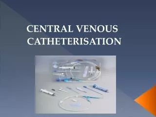
Ultrasound Guided Central Venous Cannulation (CVC). Shirley Lee MD CAEP 2008. Why Ultrasound guidance?. Traditionally, CVC mechanical complications occur up to 15% Insertion unsuccessful up to 12% Becoming standard of care. McGee and Gould, NEJM 2003; 348: 1123-1133

E N D
Ultrasound Guided Central Venous Cannulation (CVC) Shirley Lee MD CAEP 2008
Why Ultrasound guidance? • Traditionally, CVC mechanical complications occur up to 15% • Insertion unsuccessful up to 12% • Becoming standard of care McGee and Gould, NEJM 2003; 348: 1123-1133 Merrer, De Johnghe, Golliot, et al. JAMA. 286:700-7, 2001
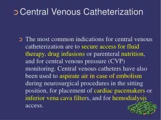
CVC Complications • Pneumothorax, arterial puncture, hematoma, malposition, increased skin punctures with bleeding complications, delay and failure to catheterize, thoracic duct injury (left sided approach), air embolism, arrhythmias, and death • Delayed complications: infection and thrombosis
Advantages • Safer • Markedly decreased pneumothorax rate • Real-time visualization of target • See needle enter target vein, avoiding adjacent arteries, nerves
Evidence • In ER: Prospective, randomized trial of 130 patients • Complication rate: 4.6% vs. 16.9% • Success rate: 93.9% vs. 78.5% • Time not significantly different Leung, Duffy, Finckh. Ann Emerg Med 2006;48(5):540-7
Evidence • ICU – Prospective,randomized trial of 900 • Success: 100% vs. 94% • Carotid puncture 1% vs. 10% • Pneumothorax 0% vs. 2.4% • Hemothorax 0% vs. 1.7% • Reduced time and number of attempts with US
Subclavian vein Transverse Sagittal
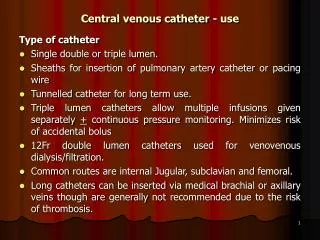
Common indications for CVC • Hemodynamic monitoring • Administration of drugs likely to induce phlebitis • Temporary cardiac pacemaker • Hemodialysis • Lack of peripheral venous access
Technique • 5-10 MHz probe - locate vein, ensure patency, then puncture blindly – but no safer than landmark technique • Real-time visualization of needle tip helps prevent pneumothorax, arterial puncture • Still need X-ray to document tip position, as catheter can still go wrong direction
Technique • Use either transverse or longitudinal orientation of ultrasound beam to needle path • Transverse supposedly easier for novice • Advantage of longitudinal: see needle through entire course • With either, you will NOT see needle tip if out of plane • Can use needle guides to help
Predictors of difficult cannulation • Emergency Placement • Obesity • Coagulopathy • Intubated • Hypotensive/Hypovolemic • Edematous patient • Known previous difficult cannulation
Static technique: Using 2-3 ultrasound planes, mark with felt pen Dynamic technique: 1 vs 2 person technique Use image as guide Observe needle throughout procedure, as it penetrates vessel Watch tip! Static vs Dynamic technique
Venous access – easiest to more challenging 1st CHOICE:Internal/External Jugular 2nd CHOICE: Femoral – easy 3rd CHOICE: Subclavian/Axillary – harder due to location, more difficult to visualize while you puncture 4th CHOICE: Cephalic/Basilic/Antecubital –
Jugular Vein • Large, easy to see, good choice • Trendelenberg, head contralaterally turned 30 degrees • Put probe transversely across vein, just superior to clavicle btn two SCM heads, just superior to clavicle • Bring needle in from laterally above probe (in same plane as transducer), aiming just slightly down to toes ~ 20-45 degrees (Posterior approach) • Watch needle well away from vein, indenting vein wall, and pop through…and know where carotid is! • Beware of anatomical variants:
Subclavian Vein • More challenging to see needle and vein at puncture site • Jugular much easier, less risk of venous stenosis, thrombosis, catheter fracture from pinch off syndrome • Place probe inferior to most lateral aspect of clavicle • Puncture axillary-subclavian junction close to clavicle
Femoral vein • Also easy • Orient transducer longitudinally, along course of vein, bring needle in from below, parallel to transducer and vein • Valsalva often helps distend vein, bigger target • Externally rotate leg to move artery more lateral
U/S CVC Pitfalls 1. Failure to identify the vein correctly 2. Failure to locate the needle in tissue
Tips • Awake patient - Check position. If patient has moved after you have landmarked, this results in a change in anatomical position of the vein • Centre vein in middle of the screen • Lighten probe pressure, as may be collapsing vein • Insert needle at sharper angle (45 - 60 degrees), to properly intersect with the vein directly under transducer
7 Steps to Success: • Use adequate gel • Confirm orientation of probe - conventionally probe head pointing to RIGHT (rub edge with finger, look at screen) • Do preliminary US - find patent target vein • Mark site (static vs direct technique) • Consider local anesthetic • Sterilize skin, sterile probe, sterile technique! • Advance the needle!
Sterile preparation of US transducer • Apply non-sterile gel to probe • Slip sterile sleeve over transducer, smooth all air bubbles away from scanning surface to prevent artifact • Secure sleeve with rubber band • Alternate: large sterile glove, with fingers folded over, palmar surface of glove is scan surface. • Sterile gel applies outer surface of glove/sterile sleeve
General Tips on CVC insertion • Be aware that more than 3 failed attempts to cannulate the vein can result in a 6 fold increase in mechanical complication. (McGee) • Aids to distinguish arterial vs. venous cannulation • A pressure transducer can be attached to the needle cannulating the vessel to confirm the presence of venous waveforms and pressure • Blood gases from the needle in the vessel can be measured and compared with known a known arterial sample
Summary • US guided procedures have a number of clinical utilities in the ED • US can improve the safety of specific procedures and success rate • Initially, can be more time consuming due to learning curve, but with practice, patience and good hand-eye coordination improve efficiency and efficacy of patient care
References • Abboud PAC and Kendall JL. Ultrasound guidance for vascular access. Emergency Clinics of North America. 22(3):749-773, 2004 • Leung, Duffy and Finckh. Ann Emerg Med 2006;48(5):540-7 • McGee DC and Gould MK. Preventing complications of central venous catheterization. NEJM. 348(12):1123-33, 2003 • Miller AH, Roth BA, Mills TJ et al. Ultrasound guidance versus the landmark technique for the placement of central venous catheters in the emergency department. Academic Emergency Medicine. 9(8) :800-805, 2002.
Procedure Video Reference • http://content.nejm.org
References • National Institute for Clinical Excellence. NICE technology appraisal guidance No.49: guidance on the use of ultrasound locating devices for placing central venous catheters. London: NICE, September 2002. www.nice.org.uk/pdf/ultrasound_49_GUIDANCE.pdf (accessed 21 Apr 2004) • The National Quality Forum. Safe Practices for Better Health Care. A consensus report.Washington, D.C. 2003. www.qualityforum.org (accessed 27 Jan 2005).