Download

1 / 65
740 likes | 1.09k Views
Advances in Laboratory TB Diagnosis. Nancy L. Wengenack , PhD, D(ABMM ). Rochester, MN February 12, 2014. Disclosures. Trek diagnostics – grant/research support. Overview. Stains for Mycobacteria Culture of Mycobacteria Molecular methods for identification of M. tuberculosis

E N D
Advances in Laboratory TB Diagnosis Nancy L. Wengenack, PhD, D(ABMM) Rochester, MN February 12, 2014
Disclosures • Trek diagnostics – grant/research support
Overview • Stains for Mycobacteria • Culture of Mycobacteria • Molecular methods for identification of M. tuberculosis • From culture • Directly from specimen • M. tuberculosis drug resistance testing • Rapid broth-based methods • Molecular markers of resistance
Mycobacterium Tuberculosis does not Stain well With the Gram Stain M. tuberculosis ghosting on Gram stain
Mycobacteria Cell Wall • Contain >60% lipid • Mycolicacids (C60-C90 fatty acids) • Waxes • Gram positive organism contains ~5% lipid • Gram negative organism contains ~20% lipid • Mycolicacid make the cell surface extremely hydrophobic and resistant to staining with basic aniline dyes or penetration by drugs Lipoarabinomannan Mycolic acid Arabinogalactan Peptidoglycan Cytoplasmicmembrane M. Tuberculosis cell wall
Mycobacterial Stains • Mycobacteria are “acid-fast” bacilli (AFB) • A complex is formed between mycolic acid and dye (carbol-fuchsin or auramine O) • The complex is resistant to destaining by mineral acids (ie, acid-fast) • So mycobacteria retain the carbol-fuchsin or auramine O stain and other bacteria do not
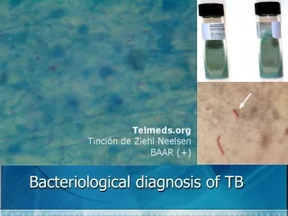
Ziehl-NeelsenStain Uses heat to help drive fuchsin stain into waxy cell wall; phenol as mordant to fix stain; (Kinyoun stain method – no heat, instead uses higher concentration of phenol and fuchsindye to aid uptake; less effective as direct stain) AFB’s stain in red; non-AFB’s stain in blue
Auramine-Rhodamine Stain 400X 1000X, oil
Other Tidbits about AFB Smears/Stains • Fite stain • Modification of ZN; often used in pathology • Uses a more mild decolorizing agent that supposedly works better for “delicate” mycobacteria like M. leprae • Tissue processing in pathology can damage the mycolic acid, sometimes making it difficult to find the AFB regardless of the stain used • LED microscopy • WHO study indicated it was superior to ZN and equivalent to fluorescence microscopy and recommended replacement of fluorescence and ZN with LED microscopy • Gaining traction in developing countries where fluorescent microscopes scarce/expensive; can run on batteries • Cannot reliably speciate using microscopy – Mtb looks like MAC which looks like M. abscessus, etc • Positive smear suggests higher likelihood of infectivity if the patient has pulmonary Mtb
Tuberculosis Cases with Pulmonary Involvement by Sputum AFB Smear ResultMinnesota, 2008-2012 n=491 Not done/unknown* 13% Positive40% Negative47% *67% of cases without sputum smear results were under 15 years of age www.health.state.mn.us/tb
Are Two AFB Smears Better than One?Yield of Serial AFB Smears
Culture of M. tuberculosis Complex Sensitivity of culture is much better than smear; only 10-100 viable organisms/mL required for positive culture Culture • Solid Medium • Egg-based – Lowenstein-Jensen (LJ); TTP ~30 days • Agar-based – Middlebrook • Rapid Broth (Liquid) Medium (FDA-cleared systems) • Reduces TTP to ~ 10 days • BACTEC MGIT (fluorimetric, BD) • VersaTREK (pressure, TREK)
Note the “rough and buff” morphology typical ofM. tuberculosis
BACTEC MGIT 960 Culture System MGIT - Mycobacterial Growth Indicator Tubes (Becton Dickinson) • fluorescent indicator in bottom of tube quenched by O2 • mycobacterial growth = O2 and fluorescence
VersaTREKSystem Mycobacterial growth causes changes in bottle headspace pressure which are detected by the instrument; sponges in bottle are supposed to provide increased surface area for growth http://www.trekds.com/products/versaTREK/mdst.asp
Tuberculosis Cases by Mycobacterial Culture ResultMinnesota, 2008-2012 Not done/unknown* 4% n=806 Negative21% Positive75% www.health.state.mn.us/tb
Traditional Methods of Identification • Historically, positive mycobacterial cultures were identified on the basis of • Colonial morphology • Growth characteristics • Biochemical testing (niacin, nitrate, pyraziniamidase) • Slow process taking up to 8 weeks • Sometimes, HPLC or GLC for cell wall constituents – generally at CDC or State Public Health Labs
Molecular Methods Allow for Rapid Identification Identification Methods for Culture Isolates
1. Nucleic Acid Hybridization Probes From culture only • No amplification step • Need lots of target nucleic acid! • Add probe with unique, complementary sequence to known species; chemiluminescent detection • Identification within 2-3 hours after growth in culture Hologic Gen-Probe AccuProbes® (nucleic acid hybridization probes) available for • M. tuberculosis complex • M. avium complex • M. gordonae • M. kansasii
Hybridization Probes Step 1 Step 2 Step 3 Step 4 DNA probe Microbiology culture plate Sonicator for15 minutes Heat at 95oc for 10 minutes Add DNAprobe reagent DNA-rRNA hybrids detected with chemiluminescent reads Lysing reagent
2. Line Probe Hybridization Assays for Mycobacteria(HainLifesciences or Innogenetics) • Genus- and species-specific probes bound to nitrocellulose membrane • DNA from lysed culture extract hybridizes to the probe for identification • GenoType Mycobacterium CM and AS • M. tuberculosis complex and 29 nontuberculous mycobacteria on 2 strips • GenoType MTBC • Differentiation of M. tuberculosis complex • GenoType MTBDR plus • M. tuberculosis complex plus wt and mutant rpoB, katG, inhA 1 Conjugate control 2 Universal control 3 Genus control 4 5 6 7 8 9 10 11 12 13 14 15 16 17 Specific probes Species may possibly befurther differentiatewith the GenoTypeMycobacterium AS For further differentiationuse the GenoTypeMycobacterium AS For further differentiationuse the GenoType MTBC M. avium M. xenopi M. gordonae M. fortuitum M. kansadii M. abscessus M. chelonae M. interjectum M. malmoense M. peregrinum M. intracellulare M. scrofulaceum Mycobacterium spec M. Marinum / M. ulcerans M. Tuberculosis complex Not approved for diagnostic use in U.S. at this time http://www.hain-lifescience.de
3. M. tuberculosis Identification by DNA Sequencing Sanger dideoxy sequencing is the current gold standard for mycobacteria identification • Various targets are useful (rpoB, hsp65, 16S rDNA gene, etc) • uses broad range primers that will amplify all mycobacteria species • hypervariable region between primers used to distinguish species Hall L et al: JCM 41:1447, 2003
Sequence Analysis • Compare the isolate sequence to known mycobacterial sequence libraries • Microseq library (AB) • Lab-specific custom library • Genbank BLAST (NCBI) • Curated, web-based database tools • Smartgene or isentio • TAT can be as fast as 8hrs after growth of the organism in culture; in our lab we run in batches of ~96 isolates • Select colonies to be sequenced in am • Pcrs in afternoon • Electrophorese overnight • Read/report next am
Advantages and Limitations of Sequencing for Identifcation of Mycobacteria Advantages • Allows for objective identification of a wide variety of mycobacteria • Next day identification after growth in culture Limitations • Labor-intensive, requires skilled, trained (dedicated) technologists • Equipment and reagent costs drive total test cost up • Results are highly dependent upon the quality of your sequence library database
4. MALDI-TOF MS – a Paradigm Shift in Microbiology • Matrix-assisted laser desorption ionization – time of flight (MALDI-TOF) mass spectrometry is changing the way we identify microbes • Already becoming the main technique used in many laboratories for bacterial and yeast identification • Mycobacteria and mold identification by MALDI-TOF MS is not far behind
Two examples of MALDI-TOF MS Instruments for Identification of Microorganisms BrukerBiotyper bioMérieuxVitek MS
MALDI-TOF MS TheelES: Clinical Microbiology Newsletter 35:155, 2013
Laboratory Workflow for MALDI-TOF MS ID of M. tuberculosis Complex after Growth in Culture BSL3 Activities Incubate room temp 10 min Bead Beat 2 minutes 10 ulloop-ful of organism Beads+500µl 70% Ethanol BSL2 Activities Decant supernatant Centrifuge 5 min Speed ac 10 min 70% Formic Acid & Acetonitrile Start to finish takes ~2 hrs for 24 samples Spot 1ul sample + 2ul of Matrix MALDI-TOF
Advantages of MALDI-TOF MS for Mycobacteria Identification • Similar work-flow regardless of organism (bacteria, yeast, mycobacteria, mold) • Cost effective and “Green” – low consumable costs • Rapid turn around time, high throughput • Automated, robust, interlaboratory reproducibility • Single colony requirement • Small footprint • Low exposure risk – sample inactivation • Adaptable – can be an open system w/ databases expandable by user
Limitations of MALDI-TOF MS for Identification of Mycobacteria • Need growth in culture • Requires pure isolate • Phase of growth, media, timing all factors • Best performance, your spectral library needs to be composed of spectra produced under comparable conditions to your everyday working practices • Databases need expansion for less common organisms • Instrument maintenance downtime (if using a single instrument) • Regulatory issues • May not be a bit slower than sequencing for slowly growing mycobacteria
Mass Spectrometry Equipment Costs • Purchase cost: ~$200,000 • Steel plates (10): ~$5,000 • Service contract (year): ~20,000 • Maintenance cost (year): ~$5,000 Remember – Mass spectrometry can also be use for identification of bacteria, mycobacteria, mouldson the same platform; next generation instruments will likely be linked with susceptibility platforms too
New Workflow for Mycobacteria ID Culture to media; wait for growth MALDI-TOF MS (same day ID) Sequencing (next day ID) If no ID
Direct Identification of M. tuberculosis Complex Without Waiting for Growth in Culture
Nucleic Acid Amplification-Based (NAA) Tests CDC recommends • NAA testing be performed on at least one (preferrably the first) respiratory specimen from each patient with suspected pulmonary TB • If it would alter case management • If it would alter TB control activities • NAA testing does not replace the need for culture
1. Mycobacterium tuberculosis Direct Test (MTD) from Hologic Gen-Probe • People frequently refer to this as the “TB probe” assay but that is not correct; this is a PCR-like amplification method • Transcription-mediated amplification of M. tuberculosis complex rRNAdirectly from respiratory specimens • Clinical specificity: 99-100% • Clinical sensitivity • Smear positive: 91-95% • Smear negative: 83-100%
Limitations of MTD test • Technically “fussy” test • Inhibition from specimen components a concern • Open PCR system so false positives due to contamination are possible • Negative does not rule out M. tuberculosis infection (still need to do a culture • Detects presence of nucleic acid but doesn’t indicate if the organism is still viable • Cross-reactions occur w/ some rare mycobacteria: M. celatum, M. terrae-like organisms, M. holsiaticum • Can be costly
2. Laboratory-Developed PCR Tests (LDTs) • Closed PCR system – reduced opportunity for false-positives • Good sensitivity and specificity but it can vary since each test developed/verified independently • Often less expensive than MTD • Some can be used on a wider variety of specimen types included smear negative specimens and formalin-fixed, paraffin-embedded tissue blocks
Example of Real-time PCR Workflow in our Laboratory Specimen or culture lysis, inactivation and processing DNA extraction PCR amplification and detection Approximate turn-around time = 4 hr
Direct Comparison of Mayo LDT PCR Assay With the GenProbe MTD Test
3. Cepheid Xpert MTB/RIF Test • WHO-endorsed • Runs on the Cepheid GeneXpert system • recently FDA-approved for respiratory specimens • Detects M. tuberculosis complex and provides information about RIF resistance www.finddiagnostics.org
XpertAccuracy for Detection of Mtb Complex Chang et al:J Infect 64:580, 2012 • Meta-analysis of 18 studies with 10,224 patients total • Pulmonary TB • Sensitivity, Smear positive disease – 90.8% • Sensitivity, Smear negative disease – 74.3% • Specificity – 98.4% • ExtrapulmonaryTB • Sensitivity – 80.4% • Specificity – 86.1% Time to diagnosis comparison • Smear microscopy = 1 day (non-specific) • Broth culture took an average of 16 days • Solid media plate cultures took an average of 20 days • Xpert – same day diagnosis
Xpert MTB/RIF and Rifampin Resistance • rpoB: gene encoding beta subunit of bacterial RNA polymerase • Mutations in an 81bp region of the rpoB gene are responsible for ~96% of RIF resistance in Mtb; also predicts MDR TB since the majority of RIF-resistant isolates will also be INH-resistant • Some false positive RIF resistance with Xpert • PPV is lower in low prevalence settings • CDC recommends reporting Xpert RIF-R as a preliminary result pending confirmation with sequencing; growth-base DST is still required
Strengths of Xpert MTB/RIF Assay • Good sensitivity and specificity for respiratory specimens • Rapid 2 hrTAT • Detect MTB and RIF resistance • Closed PCR system with low risk of cross-contamination • GeneXpert platform is multi-functional and can be used for other tests (eg, C. difficile, HIV viral load) • Simple for operators to perform • No advanced biosafety equipment needed
Weaknesses of Xpert MTB/RIF Assay • Xpert has better sensitivity than smear with respiratory specimens but a culture is still necessary • False-positive RIF resistance is possible; need to confirm RIF-resistance with sequencing • Not as sensitive or specific for extrapulmonary specimens • Expensive – need to purchase GeneXpert platform; cartridges are $65 each in E.U. and U.S.; $10 discounted price for high burden and developing countries • Need continuous electrical power and air conditioning (challenge in developing countries) • Sample storage limited to 3 days at RT, 7 days at refrigerated temps • Can’t differentiate between live and dead M. tuberculosis (can’t use to monitor treatment)
4. Line Probe Assays for Mycobacteria (HainLifesciences or Innogenetics) Conjugate control Universal control MTBC M. tuberculosis complex detection and INH/RIF resistance Specific gene probes forthe differentiation of theMycobacterium tuberculosiscomplex BCG M. microti 1 2 3 4 5 M. tuberculosis M. africanum M. Bovissspbovis M. Bovissspcaprae Conjugate control Amplification control M. Tuberculosis complex Conjugate control Amplification control M. Tuberculosis complex M. tuberculosis complex speciation rpoB Locus control rpoB wild type probe 1 rpoB wild type probe 2 rpoB wild type probe 3 rpoB wild type probe 4 rpoB wild type probe 5 rpoB wild type probe 6 rpoB wild type probe 7 rpoB wild type probe 8 rpoBmutation probe 1 rpoBmutation probe 2A rpoBmutation probe 2B rpoBmutation probe 3 rpoB Locus control rpoB wild type probe 1 rpoB wild type probe 2 rpoB wild type probe 3 rpoB wild type probe 4 rpoB wild type probe 5 rpoB wild type probe 6 rpoB wild type probe 7 rpoB wild type probe 8 rpoBmutation probe 1 rpoBmutation probe 2A rpoBmutation probe 2B rpoBmutation probe 3 katG Locus control katG wild type probe katG mutation probe katG mutation probe 2 katG Locus control katG wild type probe katG mutation probe katG mutation probe 2 Not approved for diagnostic use in the U.S. inhA Locus control inhA wild type probe 1 inhAwild type probe 2 inhA mutation probe 1 inhA mutation probe 2 inhA mutation probe 3A InhA mutation probe 3B Colored marker inhA Locus control inhA wild type probe 1 inhAwild type probe 2 inhA mutation probe 1 inhA mutation probe 2 inhA mutation probe 3A InhA mutation probe 3B Colored marker Resistance - R+I I R+I R+I R = Rifampicin I = Isoniazid http://www.hain-lifescience.de