Download
1 / 115
1.15k likes | 1.2k Views
Learn about subject and film contrast in radiography, the impact of contrast media, and how to enhance contrast for better medical imaging. Discover the types of contrast media and their administration methods.

E N D
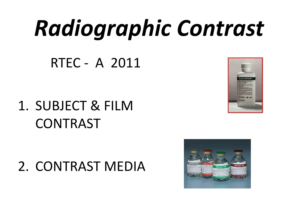
Radiographic Contrast RTEC - A 2011 • SUBJECT & FILM CONTRAST • CONTRAST MEDIA
2 types of Radiographic “Contrast” • Subject contrast • patient • Film contrast • Inherent in equipment • The BLACKS & WHITES ON THE FILM / IMAGE
Subject Contrast • Range of differences in the intensity of the x-ray beam • After it has been attenuated by the subject.
SUBJECT CONTRASTRadiographic object - influenced by • Atomic Number of object • Density of object • Thickness of object • 5 materials seen on a radiograph, • Gas/air, fat, soft tissue (muscle/organs), • bone and metals
Atomic Number • Fat = 6.46 • Water = 7.51 • Muscle = 7.64 • Bone = 12.31
TissueSubject Contrast • Atomic # of object • Density of object • Thickness of object • Higher atomic # = more attenuation • Denser = more attenuation • Thicker = more attenuation
PATHOLOGY • Pleural Effusion • Excessive fluid in lung • More dense than air
Pneumothorax • Lung collapses • No tissue in space • Easy to penetrate with x-ray photons
Radiographic Contrast influenced by: • Radiation Quality (KVP) • Film attributes • Radiographic object (Patient)
What is good contrast ? • High contrast (black and white) • Low contrast (more shades of gray)
RADIOGRAPHIC IMAGE Radiation Quality = kVp • High kVp ↑ 80 • Low contrast • Lots shades of gray • Long Scale • Little differences in adjacent structures • Low kVp ↓ 70 • High contrast • Black and White • Short Scale • Great differences in adjacent structures
Contrast changes with the use of a grid Less scatter radiation – shorter scale = “better contrast” With Grid No Grid
QUALITY – KVP • A visible change in contrast will not be seen until kVp is changed 4-12 % • kVp level change change in kVp • 30-50 kVp 4-5 % 1-3 kVp • 50-90 kVp 8-9 % 4-8 kVp • 90-130 kVp 10-12 % 9-16 kVp
Scenario • Low subject contrast in the area of interest. • You want to see the difference between muscle & fat & organs? • What can be done to attain medical information and define organ structure and function? • _____________________________________
Scenario • Low subject contrast in the area of interest. • You want to see the difference between muscle & fat & organs? • What can be done to attain medical information and define organ structure and function? • USE CONTRAST MEDIA
Purpose of Contrast Media • To enhance subject contrast or render high subject contrast • In a tissue that normally has low subject contrast. • Creates bigger differences in atomic number (z #’s)
Categories of Contrast Media Negative contrast • (AIR OR CO2) • Radiolucent • Low atomic # material • Black on film Positive contrast • (all others) • Radiopaque • High atomic # material • White on film
RADIOLUCENT - dark on image • AIR, CO2 • RADIOPAQUE - white on image • BARIUM • IODINE
AIR / CO2 Naturally seen in the LUNGS STOMACH (gas in intestines) Negative Contrast
2 BASIC TYPES OF ‘”Positive” CONTRAST MEDIA BARIUM Z# 56 KVP 90 – 120* • NON WATER SOLUABLE • GI TRACT ONLY INGESTED OR RECTALLY IODINE Z# 53 KVP BELOW 90* USUALLY 70 – 80 KvP • WATER SOLUABLE • POWDER • LIQUID • INTRAVENOUS OR • GI TRACT • OIL BASED • DUCTS /ORGANS
Positive Contrast Material INGESTED /INSTILLED • (ORALLY OR RECTALLY) • BARUIM • IODINES • GASTROGRAFIN • HYPAQUE POWDER INJECTED • IV – INTO BLOOD VESSELLS • Organs and ducts • IODINES • IONIC OR NON-IONIC • VESSELLS & ORGANS • OIL BASED • DUCTS /ORGANS ONLY
Methods of Administrationof Contrast Material • INGESTED / INSTILLED • (ORALLY OR RECTALLY) • INJECTED • IV – INTO BLOOD VESSELLS • RETROGRADE • AGAINST NORMAL FLOW (Vessels & Organs) • INTRATHECAL • Spinal canal • PARENTERAL • (IV, Intrathecal) • Injecting into bloodstream • (anything other than oral)
BARIUM BARIUM SULFATE
HISTORY OF BARIUM BaSo 4 • LEAD SUBSTRATE – TOXIC • BISMUTH SUBNITRATE – TOXIC • THORIUM – RADIOACTIVE • BARIUM SULFATE - INERT • (goes in and comes out the same – not absorbed) • NOTE SOME PATIENT MAY SHOW ALLERGY TO SUSPENSION SOL.
Barium Sulfate BaSO+ • High atomic number • Not soluble in water • Used to coat the lining of organs • Supplied in different thicknesses • Used • Esophogram, UGI, Small Bowel,Lower GI or BE
Barium Sulfate BaSO+ • Because it is not water soluble – it must be mixed in a SUSPENSION with water • FLOCCULATION – when barium clumps (separates from the water) • Barium residue in the colon can dry and cause an obstruction • Drink plenty of fluids after exam
BARIUM • MIXED IN A SUSPENSION • MUST BE SHAKEN • CHECK THE CAP (LID) FIRST !!!!!!! • SUSPENSION – sodium citrate, vegetal gums, flavoring and sweeteners to improve palatability
ADVERSE REACTIONS • SUSPENSION MAY CAUSE ALLERGY • OCG TABLETS (IODINE) ALLERGY • AFTER EXAM – MAY SOLIDIFY DIFFICULT TO EVACUATE • INCREASE FLUIDS, MILD LAXATIVE • EXTRAVASATION OF CONTRAST INTO PERITONEUM
BARIUM CONCENTRATION • DIFFERENT FOR EXAMS • W/W RATIO (weight/weight) • Mixture of barium to water – 100 g suspension • “THICK” VS “THIN” BARIUM
BARIUM “THICK & THIN” • THICK – • DOUBLE CONTRAST • THIN – • SINGLE CONTRAST
BARIUM ORAL OR RECTAL • LABELS ARE DIFFERENT • CHECK CAREFULLY BEFORE GIVING TO THE PATIENT
Palatability OF BARIUM • Chalky taste with barium sulphate/water mixture • Contain a flavoring agent, sweetners • To disguise the unpleasant taste • Thicker or thinner suspensions may be used • Many commercial preparations contain carboxymethyl cellulose (Raybar, Barosperse) 1. Which retains fluid and prevents precipitation of the barium suspension in the normal small bowel
GASTOINTESTINAL exams • BARIUM COATS LINING OF INTESTINE • SINGLE CONTRAST - BARIUM ONLY • DOUBLE CONTRAST – WITH AIR • CARBON DIOXIDE TABLETS – • FIZZIES / CRYSTALS • SODA • ROOM AIR (LOWER GI)
EXTRAVASATION • LEAKAGE THROUGH A DUCT OR VESSEL OR ORGAN INTO THE SURROUNDING TISSUE • Barium should not be given in cases of suspected perforation
Extravasation • Following a Colonoscopy with biopsy