Download
1 / 37
370 likes | 456 Views
PERFORMING RELIABLE VISUAL FIELDS NOA Fall Conference Presented by Jill Luebbert , CPOT, ABOC. What are “Visual Fields”. A test? A view of the plains of Nebraska? “the area or extent of physical space visible to the eye in a given position. How much can we see?.

E N D
PERFORMING RELIABLE VISUAL FIELDS NOA Fall Conference Presented by Jill Luebbert, CPOT, ABOC
What are “Visual Fields” • A test? • A view of the plains of Nebraska? • “the area or extent of physical space visible to the eye in a given position
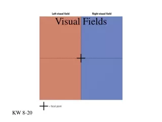
How much can we see? • Monocular visual fields usually measure • 60 degrees superior • 75 degrees inferior • 105 degrees temporal • 60 degrees nasal
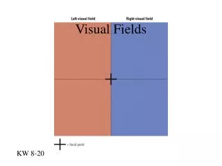
Making a map a.k.a. PerformingVisual Fields
Meridians and Quadrants • Horizontal meridian • Vertical meridian • Quadrants • Circles of eccentricity
Two methods of presenting Visual Fields • Kinetic • Static
Kinetic • Target in motion moved from non-seeing to seeing
Static • Stationary target • Threshold or suprathreshold
Terminology • What are they talking about?
Terms used • Perimetry • Visual field testing with eye located at the center of a curved instrument • Campimetry • Visual field testing eye located a specified distance from a flat surface • Scotoma • Vision entirely absent • Blind Spot (natural) • Approximately 15 degrees temporal to fixation (Optic Nerve) • Isopter • Boundary mapped for a particular stimulus size and intensity • Iso = equal opter = sight • Decibel • Relative unit, 1/10 log unit
Threshold • Every test point is evaluated by bracketing or staircase method • Suprathreshold • Target value assumed to be above threshold value for all points • False negative • Patient does not respond when a maximally bring stimulus is present at a point previously found to be normal • False positive • Patient responds when no stimulus was present
Grayscale • To be used for patient education – Represents tested points, which have been assigned value • Mean Deviation • Difference in decibels between “normal” and patient’s hill of vision • Pattern Standard Deviation • The measurement of the degree which the shape of patient’s measured “hill” of vision departs from normal • Short Term Fluctuation • A measurement of the degree of variation of threshold during the test
Types of Visual Fields • Confrontation • Harrington Flocks (Burton) Screener • Tangent Screen
Arc Perimeter Amsler Grid
Auto Perimeters • Humphrey • (Carl Zeiss Meditec) • Octopus (Haag-Steit)
Why do we perform Visual Fields? • To monitor area of vision utilized • Monitor diseases • Glaucoma • Macular Degeneration • Stroke
The technician’s responsibility • To be comfortable and knowledgeable with the instrument used • To perform accurate and reliable visual fields • To perform repeatable visual fields • To accurately gather diagnostic data • To keep the patient as comfortable and relaxed as possible • This is not a speed test
Setting up and Preparing • Data entry can be completed before the patient sits down • Choose the correct test • Set variables
Setting up • Choose the pre-determined test • Variables • Color • Fluctuation • Blind Spot Size • Test Speed
Setting Up • Enter Patient Information • Spell the name same format every time • All caps or upper and lower case • Last name first or first name fist • Middle initial with period or without period • Date of Birth • Vision Acuities • Lens Used • Pupil Size
Preparing the patient • Introduce yourself • Acknowledge them by their name • Comfortable atmosphere • Explanation of what to expect • What to see or not to see • How long to expect • Breaks??
Ready to start • Clean the instrument • New chin cover sheet • Give them the controller • Occlude non-testing eye
Positioning • Make this as comfortable as possible • Adjust the instrument height • Adjust the chin rest height • Chin and forehead firmly in place • Keep teeth clenched together • Adjust lens holder • Not too close or too far away
Ready??? • Verify the patient is in position • Give them a short test run first • All OK?? • Ready • Set • Start
Now what? • MONITOR • DO NOT LEAVE THE PATIENT • Monitor fixation • Monitor attentiveness • Is the patient staying securely to forehead rest and in chin rest
Remember • Restart if necessary • It is important to achieve reliable results • Is the patient staying attentive • Take breaks as you judge necessary • This is not a race
Finished • Are you performing the same test on the other eye? • Take a short break between • Let the patient stretch their fingers, neck • Occlude next eye • Give the patient the control • Any questions??? • Position
Keep a watchful eye • Monitor • Fixation • Patient attentiveness
Testing Complete • Save results • Return instrument control • Return testing lens • Return occluder • Print or send results to correct station • Note any observations • patient restless, etc • Escort patient to next station
“How did I do?” • Do not share results • Let the patient know they provided lots of data for the doctor to review • The doctor will visit with them regarding the results
Congratulations on performing • Thorough • Accurate • Repeatable • Visual Fields
Enjoy Nebraska Optometric Conference Experience Jill J Luebbert, CPOT, ABOC jill@jjlconsulting.com www.jjlconsulting.com Luebbert Consulting & Training