Download
1 / 38
500 likes | 1.4k Views
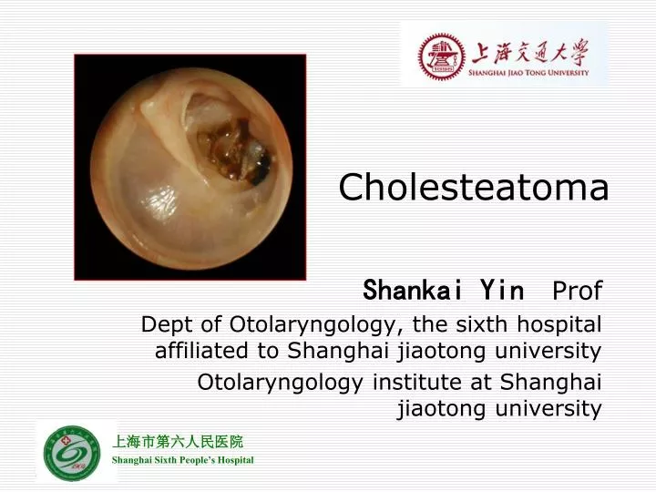
Cholesteatoma. Shankai Yin Prof Dept of Otolaryngology, the sixth hospital affiliated to Shanghai jiaotong university Otolaryngology institute at Shanghai jiaotong university. Epidemiology. Exact prevalence is unknown Incidence estimated between 3 and 12.6 per 100,000. Classification.

E N D
Cholesteatoma Shankai Yin Prof Dept of Otolaryngology, the sixth hospital affiliated to Shanghai jiaotong university Otolaryngology institute at Shanghai jiaotong university
Epidemiology • Exact prevalence is unknown • Incidence estimated between 3 and 12.6 per 100,000
Classification • Congenital • Acquired • Primary acquired (retraction pocket) • Secondary acquired
Pathogenesis • Congenital • Arise from embryonal rests of epithelial cells • Location (petrous pyramid, mastoid and middle ear cleft) • Levenson criteria • White mass medial to normal TM • Normal pars flaccida and tensa • No history of otorrhea or perforations • No prior otologic procedures • Prior bouts of otitis media not grounds for exclusion
Theories “Acquired” inclusion theory - Tos Epidermal rest theory - Teed Michael
Primary acquired • Eustachian tube dysfunction • Poor aeration of the epitympanic space • Retraction of the pars flaccida • Normal migratory pattern altered • Accumulation of keratin, enlargement of sac
Secondary acquired • Implantation – surgery, foreign body, blast injury • Metaplasia – transformation of cuboidal epithelium to squamous epithelium from chronic infection • Invasion/Migration – medial migration along permanent perforation of TM • Papillary ingrowth – intact pars flaccida, inflammation in Prussack’s space, break in the basal membrane, cords of epithelium migrate inward
Clinical manifestations • Common • Painless otorrhea • Refractory/recurrent ear infections • Conductive hearing loss • Uncommon • Vertigo/Sensorineural • Facial nerve paralysis • CNS infections • Brain herniation/CSF leak • Pneumocephalus
Diagnosis • history • Physical Examination • Otomicroscopy • Posterosuperior retraction pocket with squam • Granulation from diseased bone • Aural polyps • Pneumatic otoscopy – positive fistula response suggests erosion into labyrinth • Cultures should be obtained in infected ears
Audiology • usually conductive loss, may vary greatly; confirm with tuning forks • Imaging • CT temporal bone – definitely obtain for revision cases, complications of chronic suppurative otitis media, sensorineural hearing loss, vestibular symptoms, other complications of cholesteatoma
Imaging • Purpose • Diagnosis • Determining extent • Risk assessment • Modalities • Plain film • Computed tomography scans • Magnetic Resonance imaging
Goals of CT Imaging • Middle ear ventilation • Ossicular destruction • Epitympanum access • Mastoid cortex • Tegmen integrity • Labyrinth involvement • Facial nerve involvement • Surgical changes
CT disadvantages • Granulation tissue vs. cholesteatoma • Specific soft tissue problems • Dural involvement • Abscess • Brain herniation • Labyrinth involvement • Sigmoid sinus thrombosis • MRI needed
MR Imaging • Hypointense on T1 • Isointense to brain • Intermediate on T2 • Nonenhancing • Granulation tissue does enhance • Recurrence detection • Lesions >2mm • 90% sensitive, 100% specificity
T2 Delayed contrast T1
Differential Diagnosis • Chronic serous otitis media • Jugulotympanic paragangliomas • Cholesterol granulomas • Neurofibromas • Hemangiomas • Arachnoid cyst • Jugular bulb anomalies • Tympanosclerosis • encephalocele
Treatment Create a “dry and safe” ear
Non-surgical • Treat the Infection – Floxin Otic Drops • Decrease the inflammation – Topical steroids • Debridement of the external canal
Surgical • Atticotomy • Radical Mastoidectomy • Bondy Modified Radical (Canal wall down) mastoidectomy • Tympanoplasty and canal wall up mastoidectomy
Prognosis • Residual or recurrent cholesteatoma over 5 years – 15 to 40% • Reported to be up to 67% in the pediatric population • Close follow - up • Regular examinations needed - 6 months
Complications • Dural tear - CSF leak • Fistula of the horizontal semicircular canal (vertigo) – Up to 10% • Facial nerve injury • Injury to the sigmoid sinus / jugular bulb • Otitic Hydrocephalus • Hearing loss • 30% have conductive loss pre-operatively • Postoperatively, an additional 30% have worsening or onset of hearing loss due to extent of disease • Infection – Meningitis, Abscess, lateral sinus thrombosis – Up to 1%
Predisposing factors • Virulent organisms • Cholesteatoma and bone erosion • Presence of a congenital dehiscence (e.g.dehiscent facial canal) or a preformed pathway (e.g. skull base fracture) • Obstruction of drainage e.g. by a polyp. • Low resistance of the patient
Pathways of infection • The commonest way for extension of infection is by bone erosion due to a cholesteatoma. • Vascular extension (retrograde thrombophlebitis). • Extension along preformed pathways as – Congenital dehiscences, fracture lines, round window membrane, the labyrinth, – Dehiscences due to previous surgery.
Classification • Cranial complications • Extra-cranial complications • Intra-cranial complications
Cranial complications • Acute mastoiditis and mastoid abscesses (most common complication). • Petrositis. • Labyrinthitis. • Facial paralysis. • Osteomyelitis of the temporal bone
Extra-cranial complications • External otitis • Cervical lymphadenitis • Retropharyngeal • Parapharyngeal abscesses
Intracranial complications • Extradural abscess (commonest intracranial complication). • Subdural abscess. • Meningitis. • Brain abscess: • Temporal lobe abscess. • Cerebellar abscess. • Lateral sinus thrombosis. • Otitic hydrocephalus.
Potentially life threatening • Suppurative otorrhea, chronic headache, pain, fever – impending intracranial complication • Mental status changes, nuchal rigidity, cranial neuropathies require neurosurgical consult