Download
1 / 21
240 likes | 476 Views
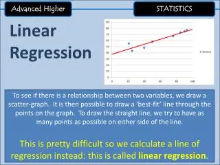
Advanced Higher Unit 3. Nuclear Magnetic Resonance Spectroscopy. NMR Spectroscopy. NMR is one of the most important analytical techniques available to an organic chemist. It uses a small sample size and is non-destructive.

E N D
Advanced HigherUnit 3 Nuclear Magnetic Resonance Spectroscopy
NMR Spectroscopy • NMR is one of the most important analytical techniques available to an organic chemist. • It uses a small sample size and is non-destructive. • A single spectrum can be used to derive a huge amount of information concerning the environment of the carbon and hydrogen atoms in an organic molecule.
Any spinning charged particle produces a magnetic field i.e. it will behave like a tiny magnet. • Any 1H nucleus will have either of two possible spin states. • The 1H nucleus is simply a single proton, therefore it behaves like a spinning proton and so produces a magnetic field. • The nuclei with one spin state will align themselves in the direction of the magnetic field while the nuclei with the other spin state will oppose the magnetic field.
Under normal conditions these magnetic fields will not interact with one another, i.e.
When a strong magnetic field is applied, these nuclei will line up along the applied field. • However for reasons best left to quantum mechanics, some of the nuclei magnetic field line up with the applied field and others line up against it. Applied Magnetic Field
These nuclei can be describes as being in a low energy state ( i.e. those aligned with the field) or a high energy state ( i.e. those aligned against the energy field)
The energy difference between the low and high energy states corresponds to the energy of electromagnetic radiation in the radio frequency (rf) range (60 MHz - 1000 MHz). • An rf pulse applied to the nuclei in the magnetic field will supply enough energy to excite (or ‘flip’) some nuclei from the low to the high energy state.
Energy High energy Energy emitted as nuclei fall back to lower state rf pulse Emitted rf radiation detected and measured Low energy • When the pulse ends the excited nuclei will flip back to the lower energy state. When this happens radiation will be emitted. • The frequency of the rf pulse must be in ‘resonance’ with the nuclei before it can be excited.
The frequency of the radiation emitted as the nuclei flip from the high energy state to the low energy state is then measured. • The intensity of the radiation emitted is very weak therefore the pulse must be applied many times and the measurements repeated and added together to build up useful results.
Why is NMR useful? • The energy difference between the low and high energy states will not be the same for the different hydrogen nuclei within a molecule. The energy difference will depend on two factors - 1) Strength of the applied field. • The stronger the field the bigger the energy difference. • This is normally kept constant during an NMR experiment.
2) The ‘environment’ of the hydrogen nuclei. • Protons aren’t the only moving charges in a molecule. • Electrons will also generate a magnetic field. • These magnetic fields ‘shield’ the hydrogen nuclei from the full effect of the applied magnetic field. • The degree of shielding, and hence the magnetic field, and therefore the energy difference, experienced by each hydrogen nuclei in a molecule will depend on the density of electron clouds of the surrounding atoms, i.e. the ‘environment’ of the hydrogen nuclei.
This means that hydrogen nuclei in different environments will emit rf radiation with differing frequencies. • By measuring the emitted frequencies and comparing them with a correlation chart (p 15 of Data Book) you can discover how many different hydrogen environments there are in a particular compound. • The area underneath the peak in an NMR spectra will also tell you how many hydrogen atoms share a particular environment.
Different NMR spectrometers apply different strengths of magnetic field and therefore give different results. • A reference standard is used in all NMR experiments so that results from one spectrometer can be compared with another. • That reference standard is Tetramethylsilane (TMS) • Any solvents used must not contain any 1H atoms. So ‘deuterated’ solvents are used, e.g. CDCl3. N.B. Deuterium (D) = 2H
A Low Resolution NMR Spectrometer along with TMS standard
The applied rf pulse contains a wide enough range of frequencies to flip nuclei in all the different environments. • The scale used to measure the emitted radiation is called the chemical shift () and has the units parts per million (ppm). • The peak associated with TMS is allocated a value of 0 (zero) on the scale.
Why TMS? • TMS has 12 equivalent hydrogen atoms (they all have the same environment) and therefore produces a sharp signal. • The signal it produces is well away from the region of signals produced by other organic hydrogen atoms.
Interpreting NMR • The number of peaks will give the number of different environments of the hydrogen atoms. NOTE The number of peaks does not give you the number of hydrogen atoms. e.g. The NMR for benzene, C6H6, will only show one peak as all the hydrogen atoms are equivalent.
The ratio of the areas of each peak will give you the number of hydrogen atoms in each environment. • The areas may be given by an integration curve, that you will have to measure using a ruler.
An NMR Spectra Methanol 1 : 3 peak ratio
The large peak can be assigned to the hydrogen atoms of the methyl (CH3-) group. • The small peak can be assigned to the hydrogen on the hydroxyl (-OH) group. • The hydroxyl hydrogen is ‘shifted’ more than the methyl hydrogen atoms.
Exercise • Now try the exercise on pages 16 and 17 of your Unit 3(d) notes.