Download
1 / 71
850 likes | 1.93k Views
Biosignaling. CH353 March 13–25, 2008. Summary. Introduction to Biosignaling Receptor-Ligand Specificity and Sensitivity Signal Transduction Themes Signaling with G Protein Coupled Receptors GPCRs stimulating or inhibiting adenylate cyclase GPCRs stimulating phospholipase C

E N D
Biosignaling CH353 March 13–25, 2008
Summary • Introduction to Biosignaling • Receptor-Ligand Specificity and Sensitivity • Signal Transduction Themes • Signaling with G Protein Coupled Receptors • GPCRs stimulating or inhibiting adenylate cyclase • GPCRs stimulating phospholipase C • GPCRs involved in sensory reception • Signaling with Receptor Enzymes • Receptors with guanylyl cyclases • Receptors with intrinsic tyrosine kinase activity • Receptors that recruit tyrosine kinases • Regulation of Cell Cycle Protein Kinases
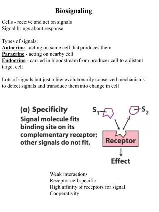
Types of Signaling Endocrine signaling • Signaling molecules act on distant target cells • hormones Paracrine signaling • Signaling molecules act on nearby target cells • neurotransmitters, growth factors, cytokines Autocrine signaling • Signaling molecules act on originating cell • tumor growth factors Juxtacrine signaling • Attached signaling molecules act on adjacent target cells • integrins, cell adhesion molecules
Signal Reception Amplification Transduction Response(s) Signal Transduction Pathways Common Elements • Receptor mediated transfer of signal inside of cell (mostly membrane receptors) • formation of receptor-ligand complex • most ligands remain outside cell • Relay and amplification of signal from receptor-ligand complex • cascades of protein and enzyme modifications and product synthesis • GTPase switch proteins, kinases and phosphatases, second messengers • Termination of signal • hydrolytic enzymes, membrane transport
Specificity of Biosignaling • Molecular complementarity between signal and receptor • multiple non-covalent interactions similar to substrate-enzyme, solute-transporter, and antigen-antibody interactions • Cell-specific expression of receptors • only cells with receptors specific for the signal can respond • Cell-specific expression of signal transduction proteins • same signal-receptor may activate or inhibit depending on other signal transduction proteins present • Cell-specific expression of effector proteins • differential response of liver, skeletal muscle and adipose cells to epinephrine depends on expressed enzymes
k1 k-1 k-1 k1 [R][L] [RL] (RT– [RL])[L] [RL] Bmax 1 + Kd /[L] Kd = = = [RL] = [Bound] [Free] [RL] [L] Bmax– [RL] Kd = = Analysis of Receptor-Ligand Interaction R receptor + L ligand RL receptor-ligand complex Total [receptor], RT = [R] + [RL] = Bmax Scatchard Plot Saturation Plot slope = – 1 / Kd Kd is [L] at ½Bmax
Saturation plot of bound receptor-ligand with increasing ligand concentration Scatchard plot for graphically measuring Kd and total number of receptors (Bmax) Analysis of Receptor-Ligand Binding
Assay of insulin binding to liver cells for measuring Kd and number of receptors per cell Increasing amounts of [125I]insulin added to liver cells Incubate at 4ºC for 1 h; separate bound and unbound [125I]insulin Curve A shows total bound insulin Control assay of [125I]insulin with 100x excess unlabeled insulin for non-specific binding (curve C) Difference is specific binding (curve B) Analysis of data indicates Kd ~ 1.4 x 10⁻⁸ M ~ 33,000 receptors/cell Measurement of Insulin Binding to Receptor
Sensitivity of Biosignaling • High affinity of ligand (signal) for its receptor • Kd for receptor-ligand > [ligand] under unstimulated conditions • Cooperativity of ligand-receptor interaction • multiple ligands to single receptor (acetylcholine) • dimerization of receptors to one ligand (cytokine) • Receptor occupancy at maximum physiological response • relatively few receptors occupied per cell for biological activity • Amplification of signal by enzymatic cascades • multiple levels of enzymes activating enzymes resulting in geometric amplification of input signal
Norepinephrine Natural Hormones and Synthetic Analogs • Norepinephrine (prohormone) • biosynthetic precursor of epinephrine • Epinephrine (hormone) • Isoproterenol (agonist) • binds to receptor and has normal biological activity • Propranolol (antagonist) • binds to receptor but has no biological activity
Biological Activity and Ligand Binding • Biological activity of Isoproterenol (IP), Epinephrine (EP) and Norepinephrine (NEP) • Binding of ligands to receptor, measured by competition assay with constant amount of [3H]alprenolol (Kd ~ 3 x 10⁻⁹ M) and increasing IP (Kd ~ 2 x 10⁻⁶ M), EP (Kd ~ 5 x 10⁻⁵ M) and NEP (Kd ~ 5 x 10⁻⁴ M)
Biological Activity and Receptor Occupancy • 50% of maximum biological activity with ~18% of receptors occupied • >80% of maximum biological activity with 50% of receptors occupied • Epinephrine levels of ~10-10 M can stimulate gluconeogenesis in liver cells, despite its relatively low binding affinity (Kd ~ 10-5 M)
Signal Transduction: Common Themes Protein and metabolite carriers involved in transducing, amplifying and transmitting signal from receptor-ligand • GTPase switch proteins (G proteins, Ras proteins) • Second messengers (cAMP, cGMP, DAG, IP3, Ca2+) • Cascades of Tyr and Ser/Thr kinases and phosphatases Clustering of receptors and signal transduction proteins • Adapter protein domains (synapses, scaffolds) • Lipid rafts (caveolin, sphingolipid, cholesterol, PIP, GPI protein) Interaction and regulation of signaling pathways • Receptor-ligand specific, cell specific • Signal integration, desensitization (receptor endocytosis)
GTPase Switch Proteins • Structure of Gsα with • GDP (inactive) and • GTP (active) • 3 switch peptides close on GTP (g phosphate) when it replaces GDP • Some require GEF (guanine nucleotide exchange factor) for activation and GAP (GTPase activating factor) for inactivation
GTPase Switch Proteins • Activation-Inactivation cycle for Ras and Ras-like proteins • Requires a guanine nucleotide exchange factor (GEF) and GTP for activation • Requires a GTPase activating protein (GAP) for inactivation • Activated G protein coupled receptors have GEF activity • Gα subunits have intrinsic GTPase activity (time delayed) • Some downstream effectors have GAP activity
Lipid Rafts and Signal Transduction • microdomains on surface of plasma membrane • segregate proteins based on attached lipid • acylated proteins in raft • prenylated proteins not • caveolin causes inward curvature forming caveolae • localized in lipid rafts / caveolae: • G-protein coupled receptors • Tyr kinase receptors (some) • not in lipid rafts: • Ras and Gg subunit (prenylated)
Signaling with G-Protein Coupled Receptors Receptors (GPCRs) • integral membrane proteins with 7 transmembrane segments • binding site for diverse ligands (hormones, odorants, tastants, light) • >907 human GPCRs (384 olfactory receptors) G Proteins • trimeric complexes of a, b and g subunits (20 Ga, 5 Gb, 12 Gg) • Ga is GTPase switch protein (GDP off / GTP on) • attached to membrane: Ga is acylated: Gg is prenylated Effectors • adenylate cyclase, phospholipase C, phosphodiesterase, channels • control levels of secondary messengers (cAMP, cGMP, DAG, IP3)
β-Adrenergic Receptor Signal Transduction Signal transduction of epinephrine depends on the type of receptor: β-adrenergic receptors stimulate adenylate cyclase α1-adrenergic receptors inhibit adenylate cyclase α2-adrenergic receptors stimulate phospholipase C
G Protein Activation Cycle Activated receptor is the guanine nucleotide exchange factor (GEF) Some effectors are GTPase activating proteins (GAP)
Interaction of Gsα and Adenylate Cyclase • Structure of adenylate cyclase 2 cytoplasmic domains (blue) bound to • forskolin (yellow) locks adenylate cyclase in active conformation • Switch helix of Gαs docks with activated adenylate cyclase • Structure of activated Gαs subunit with bound GTP (red)
Activation of Protein Kinase A with cAMP Structure of PKA catalytic subunit with bound peptide substrate R subunit domain inhibits catalytic subunit by binding to substrate site
Epinephrine Cascade • extracellular [epinephrine] > 0.1 nM (10-10 M) is sufficient for activation • intracellular [cAMP] = 1 μM (10-6 M) • PKA: GPK: GP ratio = 1: 10: 240 • PKA inactivates • glycogen synthase (GS) • phosphoprotein phosphatase (PP) • at low [cAMP]: PP is activated • PP activates GS • PP inactivates GPK and GP high cAMP: glycogenolysis low cAMP: glycogenesis GPK GP
GPCR signals with stimulatory Gsα Epinephrine (β-adrenergic) Glucagon Corticotropin (ACTH) Corticotropin-releasing hormone Histamine H2 GPCR signals with inhibitory Giα Epinephrine (α1-adrenergic) Prostaglandin E1 (PGE1) Adenosine A1 Somatostatin GPCRs with Adenylate Cyclase as Effector
Regulation of Signaling from GPCRs • Receptor-ligand affinity decreases with GTP exchange • dissociation of ligand terminates signal • GTP bound to Gα is rapidly hydrolyzed • GAP activity of effector stimulates GTPase activity • Second messenger is inactivated • cAMP phosphodiesterase hydrolyzes second messenger • Restricted localization of signaling proteins (anchoring) • A kinase associated proteins (AKAPs) anchor PKA and PDE to subcellular locations • Continuous binding of ligand to receptor is required for sustained signal transduction
Desensitization of β-Adrenergic Receptors • Continous stimulation with ligand leads to desensitization • effector kinases modify signaling and non-signaling receptors heterologous desensitization • GPCR kinases e.g. β-adrenergic receptor kinase (βARK) modify activated receptor inhibiting it homologous desensitization • arrestins e.g. β-arrestin (βarr) bind phosphorylated receptor blocking G protein signaling • Receptor-arrestin complex may be removed from membrane by endocytosis Desensitization modulates physiological response
cAMP independent pathways: βarrestin forms scaffold for alternative signaling pathways Mitogen activated protein (MAP) kinases provide signaling cascade for gene regulation Raf-1→ MEK1 → ERK1/2 → cell responses, e.g. cell proliferation, differentiation, and survival cAMP-dependent pathway: PKA translocates to nucleus and phosphorylates transcription factor CREB (cAMP response element binding) protein CREB bindings to CRE’s on DNA for initiating transcription Regulation of Gene Activity by GPCRs
Bacterial Toxins Disrupt G Protein Signaling Cholera toxin (Vibrio cholera) • transfers ADP-ribose from NAD+ to Gsα • blocks GTPase activity; Gsα is always active (bound GTP) • adenylate cyclase activation keeps [cAMP] high for days • intestinal epithelium secretes excess Cl–, HCO3– and H2O Pertussis toxin (Bordetella pertussis) • transfers ADP-ribose from NAD+ to Giα • blocks GTP exchange; Giα is always inactive (bound GDP) • adenylate cyclase not inhibited; causes high [cAMP] • pathology localized to respiratory epithelium (whooping cough)
Group Problem • Why would individuals with a recessive gene for cystic fibrosis be resistant to cholera? • How may cholera toxin and pertussis toxin be used for distinguishing which G protein is used by a receptor for signal transduction?
Fluorescent Proteins • Structure of green fluorescent protein (GFP) from jellyfish • Chromophore is autocatalytically formed by cyclizing and oxidizing SYG sequence • Site directed mutagenesis of GFP produced variety of other fluorescent proteins of different wavelengths • Combination of two FPs is basis of Fluorescence Resonance Energy Transfer (FRET) assays for protein interactions in vivo
Assay for Measuring Protein Interactions • Fluorescence Resonance Energy Transfer (FRET) uses emission of one chromophore as excitation for a second chromophore • If proteins interact excitation of 1st chromophore gives emission of 2nd • Applied to signal transduction study
Selecting for Protein Interactions Yeast 2 Hybrid System • Make construct encoding Gal4 binding domain fused to bait protein • Make cDNA library of coding regions fused to Gal4 activation domain • Transform 1st construct into yeast (select marker 1) • Transform library into transformed strain (select markers 1,2) • Select double transformants for reporter gene
GPCRs Activating Phospholipase C • Receptors: Epinephrine (α2-adrenergic), Glutamate, Histamine H1, Acetylcholine (muscarinic M1) Platelet derived growth factor, Oxytocin, Vasopressin • G Proteins: Gqa or Goa plus Gbg (various) • Effector: Stimulates Phospholipase C (β isoform) hydrolyzes phophatidylinositol 4,5-bisphosphate (PIP2) • Messengers: Diacylglycerol (DAG), inositol 1,4,5-trisphosphate (IP3), Ca2+ release • Targets: Protein kinase C (PKC) activation Calmodulin (CaM) – regulatory subunit of enzymes Ca2+/CaM-dependent protein kinases (CaM kinases) • Response: PKC has various metabolic and cell proliferation targets Sustained activation of PKC by phorbol esters interferes with normal cell growth and division (tumorigenic)
GPCR Signaling by Phospholipase C • Receptors use GqαorGoαproteins • Activated Gqα-GTP activates its effector phospholipase C (PLC) • PLC hydrolyzes PIP2 (phosphatidyl- inositol 4,5-bisphosphate) giving 2 second messengers: • diacylglycerol (DAG) and • inositol 1,4,5-trisphosphate (IP3) • IP3 opens ligand-gated Ca2+ channel on ER; cytosolic [Ca2+] ↑ • Ca2+ and DAG activates protein kinase C (PKC) on membrane • PKC phosphorylates cellular response proteins
Regulation of Cytosolic [Ca2+] • IP3-gated channels in ER release Ca2+ into cytosol • cytosolic [Ca2+] lowers affinity of gated channels for IP3 • causes oscillation in cytosolic [Ca2+] • cytosolic [Ca2+] measured using fluorescent Ca2+-binding dye • Time course of cytosolic [Ca2+] with α1-adrenergic receptor stimulation by epinephrine • high sustained Ca2+ release may be toxic
Ubiquitous regulatory protein for transducing effects of Ca2+ 4 high affinity Ca2+ binding sites (Kd ~ 10-6 M) Regulates enzymes at helical CaM binding sites; binding involves conformation change CaM is member of Ca2+ binding protein superfamily including troponin Structure of Calmodulin: binding to 4 Ca2+ binding to regulated enzyme (red helix) EF hand folding motif Calmodulin (CaM)
Proteins Regulated by Ca2+ and Calmodulin Signal Transduction Proteins • Adenylate cyclase (brain) • Ca2+/Calmodulin-dependent protein kinases (CaM kinases I-IV) • Calcineurin (phosphatase allowing nuclear translocation of NFAT) • cAMP phosphodiesterase • cAMP-gated olfactory channel • cGMP-gated Na+ and Ca2+ channels (retinal rod and cone cells) • IP3-gated Ca2+ channel • NO synthase (paracrine signaling to vascular smooth muscle cells) • Phosphoinositide 3-kinase • Protein kinase C (PKC)
Signal Transduction in Sensory Reception • Vision: • GPCRs activate phosphodiesterase that hydrolyzes cGMP • Closes cGMP-gated ion channels; hyperpolarizes membrane • Olfaction: • GPCRs activate cAMP or DAG/IP3 pathways; opens ligand-gated Ca2+ channels and Ca2+-gated Cl– channel; depolarizes membrane • Gustation: • GPCRs activates AC, PDE and/or PLC; depolarizes membrane • Ion-gated ion channels depolarize membrane • Hearing: • Mechanosensory gated ion channels depolarize membrane
Light Reception by the Eye • Photoreceptor cells • Rods: sensitive to light • Cones: less sensitive but discriminate wavelengths 3 types: red, green, blue • Rods and cones have same pigment (11-cis retinal) but different apoproteins (opsins) that shift l of activating light
Light-Induced Hyperpolarization of Rod Cells Resting cells: high [cGMP] open Na+ channels Vm = -45 mV Excited cells: low [cGMP] closed Na+ channels Vm = -75 mV
Interaction of Rhodopsin and G Protein • 3D structure of rhodopsin docked with G protein (transducin) • chromophore 11-cis retinal (blue) covalently bound to rhodopsin • analogous location for ligands • light induced change from 11-cis to all-trans alters conformation of rhodopsin activating G protein • note: palmitoylation of rhodopsin, N-term myristoylation of Ga and C-term prenylation of Gg
Activated Rhodopsin (light adapted) Rhodopsin (dark adapted) Activated Rhodopsin very high light low light high light * ATP ADP * * * rhodopsin kinase P arrestin P P P P P P arrestin no Gtα activation activation of Gtα slightly reduced Gtα activation greatly reduced Gtα activation Desensitization of Rhodopsin • Rod cells detect light over a range of 105 fold; requires adaptation • Rhodopsin kinase phosphorylates activated rhodopsin • Gradually reduces activation of Gtα • Binding of arrestin-1 to phosphorylated rhodopsin turns off receptor • Cone cells used for sight in bright light can also be desensitized
Signaling by Olfactory GPCRs • Ligands: • >1000 different odorants detected by humans • Receptors: • ~380 G protein coupled olfactory receptors (human) • broad, overlapping specificity for odorants • Signaling: • Golf activates adenylate cyclase → cAMP pathway • Gq activates phospholipase C → IP3/DAG pathway • opens ligand-gated Ca2+ channel; Ca2+ opens ion-gated Cl– channel; depolarizes membrane
Gustatory Signal Transduction • Sweet • GPCRs with Ggust activating AC, cAMP activates PKA, PKA phosphorylate (closes) K+ channel depolarizing membrane • Bitter • Ga activates cAMP-phosphodiesterase, inactivating cAMP • Gbg activates PLC, producing DAG and IP3, releasing Ca2+ • Umami • GPCRs activate cAMP-phosphodiesterase, inactivating cAMP • Salty • Na+ gated Na+ channel opens, depolarizing membrane • Sour • H+ opens gated H+ and Na+ channels, closes K+ channel, depolarizing membrane
Diverse Signals – Analogous Signaling Signal: Receptor: G Protein: Effector: Messenger: Target: