Download
1 / 42
420 likes | 433 Views
Learn about the structure and function of the integumentary system, which includes the skin, hair, nails, and glands. Discover how the skin acts as a barrier, provides sensory information, regulates body temperature, and produces important vitamins.

E N D
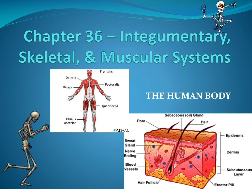
Chapter 36 – Integumentary, Skeletal, & Muscular Systems THE HUMAN BODY
A. Introduction • Humans are the most complex organisms on Earth. Our bodies are composed of trillions of _________, the smallest unit of life. These cells work together to form ______________. Cells Tissues
There are 4 types of tissues in the human body: body surfaces • Epithelial Tissue – Covers ______________; lines ________________. May contain _________ for secretions or cells with _____. • Examples includes glands, blood vessels, skin. • Connective Tissue – Is the most ________________ tissue in the body. Used as ________________ and for ____________________________. Contains a network of non-living material called a _____________. • Examples include bone, blood. Organs & vessels glands cilia abundant connectors support, transport, storage matrix
There are 4 types of tissues in the human body: electrical force movement 3. Muscle Tissue – Able to generate __________signals that create _______ and _____________. • Nerve Tissue – Specialized to generate and _______________ electrical signals to ____________________. These tissues work together as ________, which work together as ________________. transmit transfer information organs organ systems
B. Organ Systems of the Human Body Nervous • __________System – Receives, processes, & transmits information; coordinates all body systems. 2. __________ System – Regulates homeostasis with chemicals known as hormones. 3. _________ System – Supports and protects body parts. 4. ___________ System – Produces movement. 5. _______________ System – Physical barrier against pathogens, injury, dehydration. Endocrine Skeletal Muscular Integumentary
B. Organ Systems of the Human Body Circulatory 6. ___________ System – Transports O2 , CO2 , nutrients, wastes. 7. ___________ System – Responsible for exchange of O2 , CO2 • _________ System – Destroys pathogens • __________ System – Breaks down food molecules to absorbable monomers • ________ System – Washes blood; regulates blood volume • _____________ System – Produces gametes; site of embryo development in females Respiratory Immune Digestive Urinary Reproductive
II. INTEGUMENTARY SYSTEM skin • The integumentary system is composed of the ______ and its accessory structures, including _____, _______, and the ________ found in the skin. The skin is the ________ organ of the human body. hair nails glands largest
A. Function barrier against pathogens, • Protection – Provides a _______________________ ____________________________. • Sensory Information • Regulationof Body Temperature • Vitamin Production – The skin produces Vitamin ___ when exposed to __________ which is important for _________________. UV light, dehydration sunlight D strong bones
B. Structure – The skin is composed of 3 layers: 1. Epidermis – Outermost layer of skin composed of __________ tissue. There are two parts to the epidermis: a. Basal Layer – Contains cells that are actively going through ________. As new cells are produced, older cells are _______________________. These cells produce keratin, a __________________. There are also cells in the basal layer called melanocytes which produce _________ , a ___________ pigment that protects the skin from _________. epithelial mitosis pushed toward surface waterproof protein melanin dark brown UV light
die dead • b. Outer Layer – As the epidermal cells get pushed away from blood vessels, they _______, so the outermost epidermal layer consists of ______ cells filled with ________ that are eventually ________________. keratin sloughed off
connective matrix collagen elastin 2. Dermis – Inner, thicker layer of skin composed of ___________ tissue. Contains a ___________ of ________ for strength and ________ for elasticity. Structures found in the dermis include: • Blood vessels – Provide ____ and _________ to cells; remove _____. Also help to maintain a constant body temperature. Heat can be conserved when blood vessels near the surface of the skin _________, or heat can be released when blood vessels _______. b. Hair follicles - _________occurs in follicle to produce hair. Hair consists of _____ cells filled with _________. Small, ________ muscles are attached to each follicle that __________ to pull hairs upright when stimulated by __________ O2 glucose CO2 constrict dilate Mitosis dead keratin smooth contract cold, fear
protect c. Nail follicles – Produced in same manner as hair. Purpose of nails is to ________ fingertips and toetips. d. Sensory receptors - Transmit information to the ______ and ___________. e. Glands – There are two types of glands located in the dermis: 1)Oil – Produce oil to _____________ 2) Sweat - ___________ of the perspiration produced by these glands requires energy in the form of _____, which is drawn from the skin and results in cooling. brain spinal cord lubricate skin Evaporation heat
Connective fat 3. Hypodermis - ___________ tissue specialized to store ___.
C. Skin Damage & Disorders 1. Burns – Classified according to depth of damage a. First degree – Damage only to __________. Skin appears ____, but without__________. May be caused by __________________________ epidermis red blisters sun, brief contact with hot object
epidermis dermis red blisters b. Second degree – Damage through _________ to ________. Most painful of all burns. Skin is _______ with __________. May be caused by longer exposure to sun, hot object.
epidermis dermis, maybe deeper blackened, charred • c. Third degree – Destroys __________; damage extends into ____________________. Skin usually appears ________________. May be caused by _ fire, electricity, chemicals –NOT sun!__Lifethreatening._
sun exposure melanoma 2. Skin Cancer – Most important risk factor is ____________. The most serious type of skin cancer is _________.
III. SKELETAL SYSTEM, 206 The _____ bones that make up the adult skeletal system are composed of ____________ tissue with a matrix of ___________which makes them _____, and ________, which makes them hard. In embryonic development, the skeleton is first made of ___________. The process of converting cartilage to bone is known as ____________ and requires addition of _____. This process is not completed until after birth. Osteocytes, which are bone cells , embedded in the bone matrix. connective collagen tough calcium cartilage ossification Ca2+
Two other kinds of bone cells: osteoclasts-break down bone & osteoblasts-produce bone. The adult skeleton is composed entirely of bone, except for • nose, ears, discs between vertebrae
A. Functions of the Skeletal System 1. Support – Provides a framework that supports the body 2. Protection – Protects many _______ from mechanical injury 3. Movement – Movement occurs when _________ muscles attached to bones contract. organs skeletal
calcium 4. Blood Cell Formation 5. Storage a. Minerals – Storage site for ________ and ______________. b. Fat phosphorus
B. Bone Structure organ Bone is an ______ composed of living tissue. It is surrounded by a tough layer of ____________ tissue called the ___________, ________ and____________ pass through the periosteum to the bone itself. connective periosteum Nerves blood vessels
periosteum support • Compact Bone – Outer bone tissue found beneath the ____________. Dense, almost solid tissue that provides ________. 2. Spongy Bone – Less dense, porous tissue provides __________________. The spaces are filled with soft tissue called ________. There are two types of bones marrow: a. Red Marrow - Location of blood cell production including all ____ blood cells, _________, and most ______ blood cells. b. Yellow Marrow – Site of _____ storage. lightweight support marrow red platelets white fat
C. Human Skeleton Anatomy ligaments Bones are held together by __________, tough bands of ___________ tissue. The point where two bones meet is called a _____. Joints are classified according to the amount of movement possible and the appearance of the bones involved. 1. Immovable or ______ Joints - _____ movement. Example: __________________. connective joint fixed No bones of cranium
cartilage cushion the ends of bones 2. Movable Joints - Most joints are moveable. The ends of the bones that form moveable joints are covered with a thin layer of ____________ to _________________________. The space between the two bones is filled with a fluid to moisten and lubricate the joint called _________ fluid. Some examples of movable joints are: a. Ball & Socket - __________________________. Examples: _____________________ b. Hinge - __________________________. Examples: _______ c. Pivot - ____________________________. Examples: ______________________ synovial Greatest range of movement hip, shoulder Back & forth movement knee Bones twist against each other vertebrae of neck
E. Skeletal System Damage • Osteoporosis – Associated with ___________. Characterized by loss of ____________ which results in increased risk of fracture • Scoliosis - ________ curvature of the spine • Arthritis - Inflammation of the _______. Caused by wear and tear on _________ cushioning the joints older women bone mass Lateral joints cartilage
D. Human Skeleton • The human skeleton has two divisions • Axial – Forms the main ____ and includes the _______________________ ______________________________ 2. Appendicular – Contains the bones that form the _______ & ____ and includes the bones that connect them to the axial skeleton including the ________________ axis cranium, mandible, vertebrae costas, sternum, sacrum, coccyx arms legs clavicle, scapula, pelvis
IV. MUSCULAR SYSTEM The primary function of the muscular system is to produce __________. The contraction of muscle tissue requires _____, so muscles are constantly carrying out __________________ and have a large number of ______________. movement ATP cellular respiration mitochondria
A. Muscles Individual muscle cells are called muscle ______. All humans have the ______ number of fibers. Muscle bulk occurs because of ___________ of muscle fibers, not an increase in the number of muscle cells. The number of fibers that can contract at one time determine an individual’s _________. The length of the contraction time is known as ___________. fibers same thickening strength endurance
B. Muscle Types Voluntary striated 1. Skeletal Muscle - __________, _______ muscle cells that fuse together to form a _______________ muscle fiber. Muscle fibers are arranged end-to-end to produce strong contractions. If the oxygen supply to muscle cells is depleted, they can switch to _____________________ for energy production multinucleated lactic acid fermentation
Involuntary striated heart 2. Cardiac Muscle - ___________, _________ muscle cells found only in the ______, with each cell having its own nucleus. Cardiac muscle cells are arranged in chains that lattice together. When the muscle contracts, the entire lattice of cells contracts together producing a powerful contraction.
Involuntary smooth 3. Smooth Muscle - __________, ________ muscle cells. Smooth muscle contractions are slow and prolonged. Found in the _________________________________________________ digestive system, urinary bladder, blood vessels
C. Skeletal Muscle Function connective tendons 1. Skeletal muscles are attached to bones by tough, __________ tissue called ____________. Every muscle has at least 2 tendons, each attached to a different bone: a.origin – muscle attachment site(s) that ______________ b.insertion – bone that is __________________. For example, the _____________ has ________ attaching it to the ________ and the________ The origin is the ________ and the insertion is the _______. does not move moved by contraction biceps brachii tendons scapula radius scapula radius
appendicular 2. Skeletal muscles attached to the bones of the _____________ skeleton work in opposing pairs. a. flexor – muscle that causes limb to ______ at _____. b. extensor – muscle that causes limb to __________ at ______. For example, contraction of the biceps brachii ______ the arm and contraction of the triceps brachii ____________ the arm. bend joint straighten joint bends straightens
D. Muscle Contraction • Muscle fibers in skeletal muscles are composed of smaller structures called myofibrils. Each myofibril is made up of even smaller structures called filaments. • The striations in skeletal muscle cells are formed by an alternation pattern of thick and thin filaments. - The thick filaments contain a protein called myosin. - The thin filaments are made up mainly of a protein called actin. • The filaments are arranged along the muscle fiber in units called sarcomeres, separated by regions called z-lines.
D. Muscle Contraction • Sliding Filament Model of Muscle Contraction- When a muscle contracts, the thin filament (actin) in the muscle fiber slides over the thick filament (myosin), shortening the sarcomere. - Thick myosin filament forms a cross-bridge with the thin actin filament. - Powered by ATP, cross-bridge changes shape and pulls the actin filament toward the center of the sarcomere. • This happens in every sarcomere within a muscle fiber at one time so the entire fiber is shortened and the muscle contracts.
Control of Muscle Contraction • The impulses from motor neurons control the contraction of skeletal muscle fibers. • The neuromuscular junction is the point of contact between a motor neuron and a skeletal muscle cell. • The impulse in the muscle fiber begins with the release of the neurotransmitter acetylcholine from the motor neuron. This sets off a series of events that allow actin and myosin filaments to interact. • A muscle cell remains contracted until an enzyme produced at the axon terminal destroys any remaining acetylcholine.