Download
1 / 25
250 likes | 411 Views
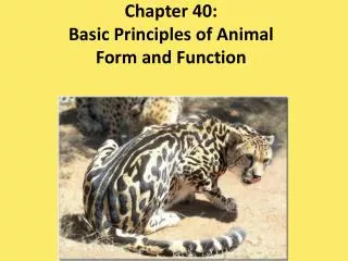
Chapter 32 – Animal Diversity. 32.1 – Animals are multicellular, heterotrophic eukaryotes with tissues that develop from embryonic layers. Animals have the following characteristics: Multicellular heterotrophs Most have muscle & nervous tissue

E N D
32.1 – Animals are multicellular, heterotrophic eukaryotes with tissues that develop from embryonic layers • Animals have the following characteristics: • Multicellular heterotrophs • Most have muscle & nervous tissue • Most reproduce sexually, with a flagellated sperm & a large egg which unite to form a diploid ZYGOTE • The diploid stage dominates the life cycle • 1.3 million living species
Vocab • Zygote • Fertilized egg • Cleavages • Successive mitotic cell divisions without cell growth between cycles • Blastula • Hollow ball of cells surrounding a cavity called the blastocoel
Gastrula • As the blastula is “punched in”, the embryonic tissue layers will form • Ectoderm • The outer tissue layer • Endoderm • The inner tissue layer
Blastopore • Opening into the gastrula • Becomes the mouth in protosomes • Becomes the anus in deuterostomes • Archenteron • Blind pouch formed by gastrulation
Some animals have larvae • Immature form distinct from the adult stage they will undergo metamorphosis • Animals share HOX GENES • Unique family of genes that play important roles in development • Can produce a wide diversity of animal morphology
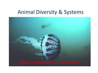
32.3 – Animals & Their “Body Plans” • Symmetry • None (sponges) • Radial • Bilateral
Radial occurs in: • Jellyfish • Any cut through the central axis would produce mirror images
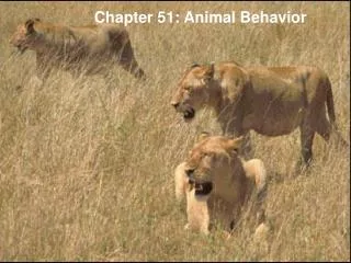
Bilateral occurs in • - Lobsters • Humans • Have a right & left side • Single cut would divide the animal into 2 mirror image halves • Dorsal side (back) • Ventral side (belly) • Anterior (head) • Posterior (tail) • Cephalization
Tissues • Animal body plans vary according to the organization of the animal’s tissues • Tissues are collections of specialized cells isolated from other tissues by membranous layers • During development, three germ layers give rise to the tissues and organs of the animal embryo
Ectoderm is the germ layer covering the embryo’s surface • Endoderm is the innermost germ layer and lines the developing digestive tube, called the archenteron • Mesoderm is the middle layer
Diploblastic animals have ectoderm and endoderm • Triploblastic animals also have an intervening mesoderm layer; these include all bilaterians
Body Cavities • Most triploblastic animals possess a body cavity • 3 types: • 1) A coelomate possesses a true body cavity • Derived from mesoderm • Filled with fluid • Separates an animal’s digestive tract from the outer wall • Earthworms
2) Pseudocoelomate • Triploblastic animals • Cavity formed from mesoderm & endoderm • Roundworms
3) Acoelomates • No cavities between alimentary canal & outer wall of body • Flatworms
Functions of Body Cavities • 1) Cushion suspended organs • 2) Act as a hydrostatic skeleton • 3) Enable internal organs to grow & move independently
Protostome & Deuterostome Development • 3 major differences: • 1) Cleavage • 2) Coelom formation • 3) Fate of the blastopore
Cleavage: P = begins with spiral, determinate cleavage D = radial, indeterminate cleavage
Coelom Formation: Begins in the gastrula stage P = coelom forms from splits in the mesoderm D = coelom forms from mesodermal outpocketings of the archenteron
Fate of the Blastospore: P = mouth forms from the blastopore D = mouth forms from a secondary opening