Download
1 / 1

E N D
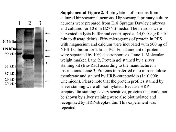
Supplemental Figure 2. Biotinylation of proteins from cultured hippocampal neurons. Hippocampal primary culture neurons were prepared from E18 Sprague Dawley embryos and cultured for 10 d in B27NB media. The neurons were harvested in lysis buffer and centrifuged at 14,000 × g for 10 min to discard debris. Fifty micrograms of protein in PBS with magnesium and calcium were incubated with 500 ng of NHS-LC-biotin for 2 hr at 4oC. Equal amount of proteins were separated by 10% electrophoresis. Lane 1, Molecular weight marker. Lane 2, Protein gel stained by a silver staining kit (Bio-Rad) according to the manufacturer’s instructions. Lane 3, Proteins transferred onto nitrocellulose membrane and stained by HRP--streptavidin (1:10,000; Chemicon). Please note that the protein profiles stained by silver staining were all biotinylated. Because HRP-streptavidin staining is very sensitive, proteins that could not be shown by silver staining were also biotinylated and recognized by HRP-streptavidin. This experiment was repeated. 1 2 3 207 kDa 119 kDa 99 kDa 57 kDa 37 kDa 29 kDa 20 kDa