Download
1 / 58
630 likes | 706 Views
Learn about radioactive decay, imaging, counting, and therapy in nuclear medicine. Understand the atom, stability of nuclei, types of decay, activity measurement, and more.

E N D
INTRODUCTION TO NUCLEAR MEDICINE Türkay TOKLU, Ph.D. Physics Engineer
Contents • Radioactive Decay • What is Nuclear Medicine? • Imaging in Nuclear Medicine • Counting in Nuclear Medicine • Therapy in Nuclear Medicine
The Atom • Nucleus • Consists of Protons (p) & Neutrons (n) • Protons are positively charged • Includes Z protons (Atomic Number) • Neutrons are uncharged • Atomic Mass (A) = p+n • Electrons • Electrons (e) are negatively charged • Z electrons turn around neutral atom
The Atom p+n=A (Atomic Mass) Charge C 14 +1 6 8 p=Z (Atomic Number) n Symbol of Atom Isotopes: Atoms with the same number of protons and different number of neutrons
Stability of a Nucleus • Stability of a nucleus depends on electrostatic and nuclear forces between protons and neutrons in nucleus. • There is no certain rule that determines which isotope will be unstable. • Natural or artificial unstable isotopes will undergo a decay process.
Radioactive Decay • It is impossible to know at what time a certain radioactive nucleuswill decay. It is, however, possible to determine the probability of decay in a certain time. • In a sample of N0 nuclei, the number of decays per unit time is then: : Decay constant t: Time N(t): Number of radioactive nuclei after time t
Half-Life, T1/2 • It is the time to remain half of initial number of radioactive nuclei of a certain isotope.
Half-Life, T1/2 IsotopeHalf-Life F-18 110 m Tc-99m 6 h I-131 8 d Na-22 2,6 y Cs-137 30 y Ra-226 1600 y K-40 1,3x10⁹ y U-238 4,4x10⁹ y Th-232 1,4x10¹⁰ y
Types of Decay • Fission • The nucleus is divided into two parts, fission fragments, and3-4 neutrons. • Examples: Cf-252 (spontaneous), U-235 (induced) • Alpha decay • The nucleus emits an a-particle (4He+2). • Examples: Ra-226, Rn-222 • Beta decay • Too many neutrons results in b-decay. n0→p++e- • Examples:H-3, C-14, I-131. • Too many protons results in b+ decayp+→ n0+e+ • Examples: O-16, F-18 • or electron capture (EC). p+ + e-→n0 • Examples: I-125, Tl-201
Types of Decay • Gamma Decay • Positron Annihilation g (364 keV) g (511 keV) 1800 g (511 keV)
Ionization (Excitation) Energy
Deexcitation Characteristic X-ray
Electromagnetic Spectrum Non-Ionizing radiation Ionizing radiation
Activity • The number of decaying nuclei per unit time. • SI unit: • Becquerel (Bq): 1 disintegration per second • Special unit: • Curie (Ci): 3,7x10¹⁰ disintegration per second (number of disintegration per second of 1 g radium) • 1 Ci = 3,7x10¹⁰ Bq = 37 GBq • Activity used in Nuclear Medicine: • For imaging: 1-30 mCi • For treatment: 100-250 mCi
Equivalent and Effective Dose • Is the measure of radiation damage to human body. • With the help of this quantity radiation effects to human can be evaluated. • SI unit: • Sievert (Sv) • Special unit: • rem • 1 Sv = 100 rem
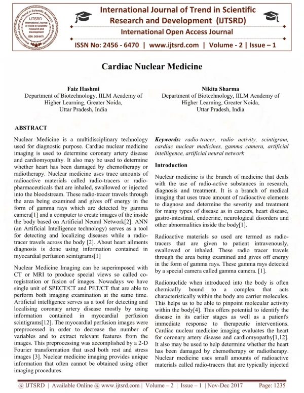
What is Nuclear Medicine? • Science of diagnosis and therapy with unsealed radioactive sources usually in liquid form. • Radioactive source is given to the patient by I.V., ingestion, drinking or breathing. • Most frequently, beta particles are used for treatment and gamma-rays are used for counting and imaging purposes. • Unlike Radiology, in Nuclear Medicine patient is the radiation source.
What is Nuclear Medicine? • Because of the chemical properties, some radioactive isotopes perfuse on certain organs. • I-131 in thyroid • Tl-201 in left ventricle of myocardium • To target other radioactive isotopes to specific organs, non-radioactive chemicals are used. • Most frequently this compounds (radiopharmaceuticals) are prepared on Nuclear Medicine departments.
Radiopharmaceuticals Radionuclide Pharmaceutical Organ Parameter + HMPAO Labeled Leukocytes Infectious Diseases + colloid Liver Reticuloendo-thelial System Tc-99m + MAA Lungs Regional Perfusion + DTPA Kidneys Kidney Function A total of 31 different radiopharmaceuticals based on Tc-99m are listed for imaging and functional studies
Imaging in Nuclear Medicine
Nuclear Medicine Imaging • Nuclear Medicine imaging detects functional (not anatomical) properties of human tissues. • The imaging is done by tracing the distribution of radiopharmaceuticals within the body with a Gamma Camera or PET system.
Gamma Camera Scintillation Crystal Collimator PMTs System electronics Gamma-ray form patient Gamma-ray perpendicular to crystal Scintillation light PMT C PMT A PMT B
Bone Scintigraphy normal pathologic
Cerebral Bloodflow Alzheimers disease normal
Myocardial Perfusion SPECT Stress Rest
Tomographic Slices of the Heart Short axis Vertical long axis Horizontal long axis
PET (Positron Emission Tomography) • Most commonly, radioactive isotopes of biological atoms, which have short half-life, are positron emitters. This fact led development of PET scanners. • PET depends on coincidence detection of gamma ray pairs produced by annihilation of a positron. • PET scanners use scintillation detectors in ring orientation.
Gated Imaging Gated Non-Gated
Gas-Filled Detectors • Measures the ion pairs produced by radiation in a gas-filled medium. Electrometer + 1234 HV - Gas The response is proportional to ionization rate (activity, exposure rate) Negative ion (e-) Positive ion