Download
1 / 22
300 likes | 1.01k Views
Mechanisms of female reproductive toxicity. Helena Taskinen Finnish Institute of Occupational Health. Critical points of female fertility. libido, sexual behaviour oogenesis hormonal function fertilization transportation implantation. Developmental toxicity.

E N D
Mechanisms of female reproductive toxicity Helena Taskinen Finnish Institute of Occupational Health
Critical points of female fertility • libido, sexual behaviour • oogenesis • hormonal function • fertilization • transportation • implantation
Developmental toxicity • gameto-, embryo- and fetotoxicity • abortion, stillbirth • teratogenic effetcs • intrauterine growth retardation • functional defects • impaired mental and physical postnatal development up to puberty • childhood cancer
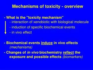
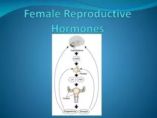
Hypothalamo-pituitary-ovarian axis • Hypothalamus: hypothalamic-releasing factor, gonadotropin-releasing hormone • Pituitary: Gonadotropin-releasing hormone, gonadotropins: follicle stimulating hormone (FSH) and luteinizing hormone (LH) • Ovary: estrogen and progesterone • Agents that disturb the axis can disrupt ovarian function
Mechanisms of toxins 1 • Direct acting toxins: structurally similar or chemically reactive • Direct damage to cells, organelles, DNA/RNA, enzymatic and biochemical pathways • alkylating compunds, metals (boron, cadmium, lead, mercury) and ionizing radiation
Indirect toxins: metabolic activation produces reactive intermediates: • cyclophosphamide, DDT, PAH, dibromochloropropane
Mechanisms of toxins 2 • Hormone agonists or antagonists • oral contraceptives, DDT, methoxychlor, polychlorinated biphenyls, polybrominated biphenyls, organochlorine pesticides • Cellular (oocyte) death: necrosis • pesticides, PAH in cigarette smoke, chemotherapeutic agents, ionizing radiation, nitrosamines, lead, mercury, cadmium, 4-vinylcyclohexene
Mechanisms of toxins 4 • Apoptosis, programmed cellular death • is preceded by activation of calcium/ magnesium-dependent endonuclease enzyme • change in the cellular environment • hyperthermia and radiation can trigger • also a physiological form of cell death • poorly understood, toxins possible, e.g. chemotherapeutics cisplatin and vinblastine
Oocyte toxicants • Polycyclic aromatic hydrocarbons can • destroy primordial follicles • cause ovarian tumors • induce chromosomal aberrations in oocyte meiosis • Busulfan and antineoplastic agents can • destroy primordial germ cells or developing follicles, and mutate preovulatory follicles
Toxicants 2 • DDT and diethylstilbestrol (DES), estrogenic compounds, suppress ovarian progesterone production • General anesthesia during periovulatory period lowers progesterone levels • Benzo(a)pyrene in cigarette smoke inhibits corpora lutea formation and thus progesterone production
Toxicants 3 • The hypothalamo-pituitary unit is disturbed by • anesthetics, stimulants, analgetics, hallucinogens, marihuana, morphine, cocaine • estrogenic chemicals, e.g. diethylstilbestrol (DES)
Toxicity of diethylstilbestrol • a synthetic estrogen, used to prevent spontaneous abortions in 1938-1971 • proven ineffective in later studies! • mutagenic and carcinogenic effects mediated through production of reactive metabolites, DNA adducts • clear cell vaginal carcinoma in daughters • 18 % of offspring (f) abnormal of the cervix
Cadmium (Cd) • Structural similarity with zinc - Cd can displace zinc in zinc-dependent enzymes • in rats: follicular atresia, changes in uterine microcirculation; decreased uterine, ovarian and pituitary weights
Developmental abnormalities • Major malformation at birth among 3 % • Problems of developmental origin among 6 -7 % by 1 year of age • Among 12 - 14 % by school age
Causes of developmental abnormalities • 20 - 28 % familial genetic defect • 10 - 3 % external exposure (environmental, drugs, nutritional) • 0 - 23% multifactorial cause • 70 - 43 % unknown cause (Wilson 1977; Nelson and Holmes 1989)
Species differences • Mammalian embryogenesis and fetal development relatively similar among all species • Differences btw. species due to differences in xenobiotic absorption and metabolism • e.g. thalidomide not soluble in rat blood - no teratogenecity in tests! When solubility was increased, teratogenic in low doses
Examples of agents causing toxic effects early in the development • Ionizing radiation • Methylnitrosourea • Medroxyprogesterone acetate • Nickel chloride • Ethylene oxide • Nitrous oxide • Isoflurane
Placenta • Provides nutrients, gas exchange and hormones for maintenance of pregnancy • Placenta is a liver, kidney, lung, ovary, pituitary and hypothalamus in one organ! • Acts as a barrier for toxicants, metabolizes them into less or more detrimental compounds
Cadmium and placenta • Cadmium induces placental necrosis at lower doses than renal toxicity • deposited in placenta, little into fetus • blocks nutrient and blood flow: growth retardation, fetal death • interferes with zinc • responsible for the growth retardation caused by smoking
Other effects on placenta • Cholinergic system regulates amino acid transport in the placenta • Nicotine, carbon monoxide, cyanide, nitrites (all present in cigarette smoke) inhibit amino acid uptake by placenta by blocking the cholinergic receptor • Risks: preeclampsia, growth retardation, premature delivery, and perinatal mortality
2-methoxyethanol (2-ME) & 2-ethoxyethanol (2-EE) and their acetates • alcohol and aldehyde dehydrogenase enzymes active; if inhibited with 4-methylpyrazole, no malformations • Teratogenic alcoxy acid metabolites: • 2-methoxyacetaldehyde and methoxy acetic acid from 2-ME • ethoxyacetaldehyde and ethoxyacetic acid from 2-EE
Heavy work • Intraabdominal pressure rises, decreases intrauterine blood flow • Growth retardation • In women 17 % fat needed for menstruation; 22 % for fertility • hypoestrogenism • In men <5 % body fat decreases testosterone and prolactin in the serum